Mitochondria supply ATP, but their protein machinery also supports central carbon metabolism, fatty-acid oxidation, calcium handling, apoptosis, and cellular stress responses. These proteins occupy distinct regions of the outer membrane, intermembrane space, inner membrane and cristae, and matrix. Human mitochondrial DNA encodes 13 oxidative-phosphorylation subunits, while most mitochondrial proteins are synthesized in the cytosol and imported into the organelle. MitoCarta3.0 catalogs 1,136 high-confidence human mitochondrial genes, although their abundance and turnover vary across tissues and physiological states (Rath et al., 2021; Morgenstern et al., 2021). This article provides an overview of mitochondrial compartments and markers, protein import and assembly, quality-control pathways, disease-related remodeling, and proteomics strategies for characterizing mitochondrial proteins.
1. Mitochondrial Compartments and Marker Proteins
Functional proteins and experimental markers should not be treated as the same category. Functional proteins describe the biology of a mitochondrial compartment, whereas markers are selected to assess localization, mitochondrial enrichment, or sample purity. Some proteins can serve both purposes, but functional relevance alone does not make a protein a reliable purity marker (Rath et al., 2021; Baker et al., 2024).
1.1 Membrane Compartments: Import, Exchange, OXPHOS, and Architecture
At the cytosolic boundary, TOMM20/22/40 recognize and translocate precursors, VDAC1-3 mediate metabolite exchange, MFN1/2 participate in fusion, and MFF/FIS1 contribute to fission control. Selected BCL2-family proteins regulate apoptosis and membrane permeability. The intermembrane space contains CYCS for electron transfer, CHCHD4 for oxidative folding, and DIABLO/SMAC, HTRA2, and AIFM1 for stress or death signaling. These proteins describe compartment function, but they are not interchangeable as purity controls.
The inner membrane houses respiratory-chain complexes, ATP synthase, SLC25 carriers, and the TIMM17/22/23 import machinery. OPA1 and MICOS shape cristae and help organize respiratory complexes. COX4I1, UQCRC2, and ATP5F1A report on different complexes, so their changes should not be read as equivalent. Coverage is also uneven: hydrophobic inner-membrane proteins are more difficult to recover and quantify than soluble matrix proteins.
Topology and release state can change how membrane-associated proteins should be read. CYCS and AIFM1 may relocate during cell-death signaling, and proteins from ER-mitochondria or lysosome-mitochondria contact sites can co-purify with mitochondrial fractions. These proteins should not automatically be dismissed as contaminants; their normal orientation, interaction partners, and response to the experimental condition provide the necessary context.
1.2 Matrix Proteins and Experimental Marker Interpretation
The matrix contains enzymes for the TCA cycle, beta-oxidation, and amino-acid metabolism, as well as TFAM, POLG, mitochondrial ribosomal proteins, and proteostasis factors including HSPD1, HSPA9, LONP1, and CLPP. CS, HSPD1, and HSPA9 are often used as matrix markers, but the choice should fit the sample and the question. Their abundance does not directly report enzyme activity or metabolic flux.
Matrix proteins work in linked modules that respond differently to nutrient supply and tissue demand. Pyruvate use, one-carbon metabolism, the TCA cycle, beta-oxidation, mtDNA maintenance, and mitochondrial translation may therefore change independently. For example, altered citrate synthase or HSPD1 can reflect a shift in mitochondrial content or stress without implying a proportional change in pathway activity.
Table 1. Mitochondrial Compartment Proteins and Experimental Markers
| Compartment | Representative functional proteins | Common experimental markers | Major functions and interpretation notes |
|---|---|---|---|
| Outer membrane | TOMM20/22/40; VDAC1-3; MFN1/2; MFF; FIS1; selected BCL2-family proteins | TOMM20; TOMM40; VDAC1 | Protein import, metabolite exchange, dynamics, and apoptosis; functional proteins alone are insufficient for purity assessment. |
| Intermembrane space | CYCS; CHCHD4; DIABLO/SMAC; HTRA2; AIFM1 (inner-membrane-associated, IMS-facing) | No universal single marker; CYCS may be used with release-state controls | Electron transfer, oxidative folding, and death signaling. Interpret localization with membrane association and release state. |
| Inner membrane / cristae | Complex I-IV; ATP synthase; TIMM17/22/23; SLC25 family; OPA1; MICOS | COX4I1; UQCRC2; ATP5F1A | OXPHOS, import, transport, and crista organization. Hydrophobicity and complex assembly affect MS coverage. |
| Matrix | CS; IDH2; MDH2; PDHA1; ACAD family; TFAM; POLG; MRPL/MRPS; HSPD1/HSPA9 | CS; HSPD1; HSPA9 | TCA cycle, beta-oxidation, mtDNA maintenance, translation, and proteostasis; activity and flux require separate assays. |
Assess mitochondrial enrichment with several positive markers from different compartments, together with negative markers for likely co-isolated organelles. Morphology or functional testing can strengthen the assessment when material is available.
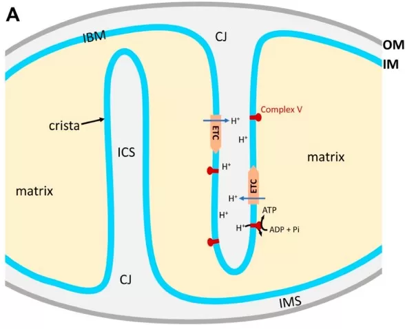
Figure 1. Structural organization of mitochondrial compartments. Major membrane regions and the matrix define distinct environments for mitochondrial proteins. Adapted from Figure 1A in Lee and Yoon (2018), Antioxidants, 7(12), 186, under CC BY 4.0. Cropped to panel A and resized; no other changes were made.
2. Mitochondrial Protein Import and Complex Assembly
Maintaining this compartmental layout requires precise targeting, translocation, processing, membrane insertion, and assembly. A defect at any step may leave precursors in the cytosol or generate incomplete complexes even when total protein abundance changes only modestly.
2.1 Targeting, Import, and Compartment-Specific Sorting
Most nuclear-encoded mitochondrial proteins are synthesized on cytosolic ribosomes and carry either an N-terminal presequence or an internal targeting signal. Cytosolic chaperones keep precursors in an import-competent state, and the TOM complex forms the main entry point at the outer membrane. Import efficiency depends on precursor folding, receptor availability, membrane potential, and coordination with downstream translocases (Bogorodskiy et al., 2021; Song et al., 2021).
Once a precursor passes through TOM, its route depends on its final destination. TIM23 moves many presequence-containing proteins into the matrix or inner membrane; TIM22 handles multi-pass carriers; MIA supports oxidative folding in the intermembrane space; SAM assembles beta-barrel outer-membrane proteins; and OXA1 inserts proteins from the matrix side of the inner membrane (Sim et al., 2023).
Import also requires energy and protein processing. The inner-membrane potential drives many TIM23 substrates, while matrix chaperones assist translocation and folding. Mitochondrial processing peptidase (MPP) removes targeting sequences from many precursors before they are stabilized, inserted into a membrane, or incorporated into a complex. Failure at these stages can cause precursor accumulation without a large change in the total protein signal.
2.2 Mitonuclear Coordination and Respiratory Complex Assembly
Respiratory complexes bring together nuclear- and mitochondrial-encoded subunits, assembly factors, and cofactors. Mature complexes form only when subunits arrive in the correct stoichiometry and sequence. Complexome profiling in yeast has captured dynamic import and respiratory-chain intermediates (Schulte et al., 2023). Although the dataset is not human-specific, it shows why a single altered subunit is weak evidence for an assembly defect.
OXPHOS subunits are synthesized in separate compartments and assembled through ordered intermediates. Conventional proteomics may show stable levels of individual subunits even when assembly factors, cofactors, or supercomplex organization have changed. Native-complex methods, complexome profiling, and targeted biochemical assays can reveal these defects more directly.
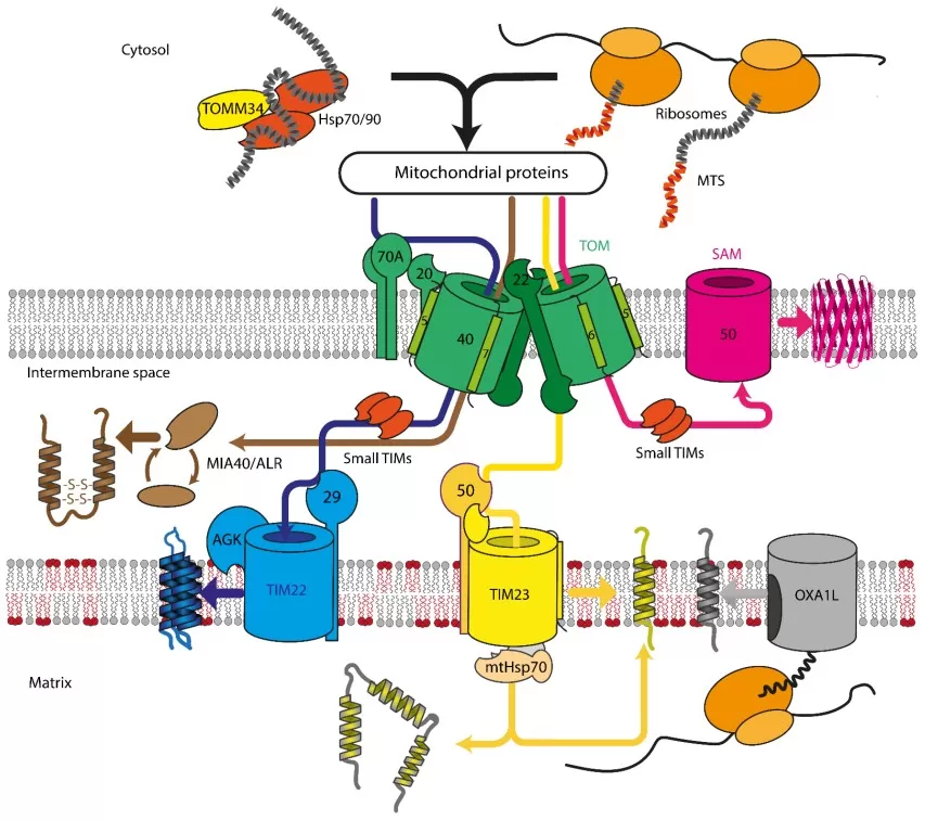
Figure 2. Major mitochondrial protein import and sorting pathways. TOM-linked pathways direct precursors to the outer membrane, intermembrane space, inner membrane, or matrix. Reproduced from Figure 1 in Bogorodskiy et al. (2021), Cells, 10(12), 3528, under CC BY 4.0; resized without content changes.
3. Mitochondrial Proteostasis and Quality Control Networks
Mitochondrial quality control operates at several scales: imported proteins are folded or degraded, damaged membrane regions are reorganized, selected cargo can be removed, and severely impaired organelles may be turned over. No single marker captures all of these processes.
3.1 Protein Folding, Proteolysis, and Import Surveillance
HSPA9/mtHSP70 and HSPD1/HSP60 assist folding after import. LONP1 and CLPP act in the matrix, whereas AFG3L2-SPG7, YME1L1, and OMA1 survey the inner membrane. The cytosolic ubiquitin-proteasome system also removes selected outer-membrane proteins, precursors that fail to enter mitochondria, and some import-stalled substrates (Song et al., 2021).
Increased chaperone or protease abundance often reflects a compensatory response to stress. UPRmt activation likewise shows that mitochondrial stress has been sensed, but it does not establish whether damaged proteins were successfully refolded or removed. Substrate accumulation, complex integrity, and functional measurements help distinguish effective adaptation from unresolved proteostasis defects.
Quality-control responses can also begin outside the organelle. Mistargeted or import-stalled precursors engage cytosolic surveillance, while mitochondrial stress can alter nuclear transcription and proteasome activity. A short-lived response may restore protein balance; persistent activation is more consistent with continuing import or folding stress.
3.2 Mitochondrial Dynamics, Mitophagy, and Turnover
MFN1/2 and OPA1 support fusion and content mixing, whereas DNM1L/DRP1 and MFF promote fission and help segregate damaged regions. OPA1 and MICOS also preserve crista architecture. Mitochondria-derived vesicles can export selected proteins or lipids without removing the entire organelle. Fission may prepare a damaged segment for repair or clearance, but it is not itself mitophagy (Giacomello et al., 2020; Jadiya & Tomar, 2020).
When local repair is insufficient, PINK1-Parkin (PRKN) signaling or receptor pathways involving BNIP3, BNIP3L/NIX, and FUNDC1 can direct mitochondria toward lysosomal degradation. PPARGC1A/PGC-1alpha, NRF1, GABPA/NRF-2, and TFAM support replacement through biogenesis; GABPA/NRF-2 is distinct from the oxidative-stress regulator NFE2L2/NRF2. Static levels of PINK1, Parkin, LC3, or biogenesis proteins cannot measure turnover or mitophagy flux. Dynamic reporters, lysosomal inhibition, imaging, or functional assays are required (Picca et al., 2023).
Mitophagy-related signaling can rise during efficient clearance, after an increase in mitochondrial damage, or when degradation is blocked at the lysosome. Biogenesis-associated proteins pose a similar interpretive problem because their increase does not confirm that newly formed mitochondria are functional. Time-course and perturbation experiments can distinguish these possibilities more reliably than a single proteomic measurement.
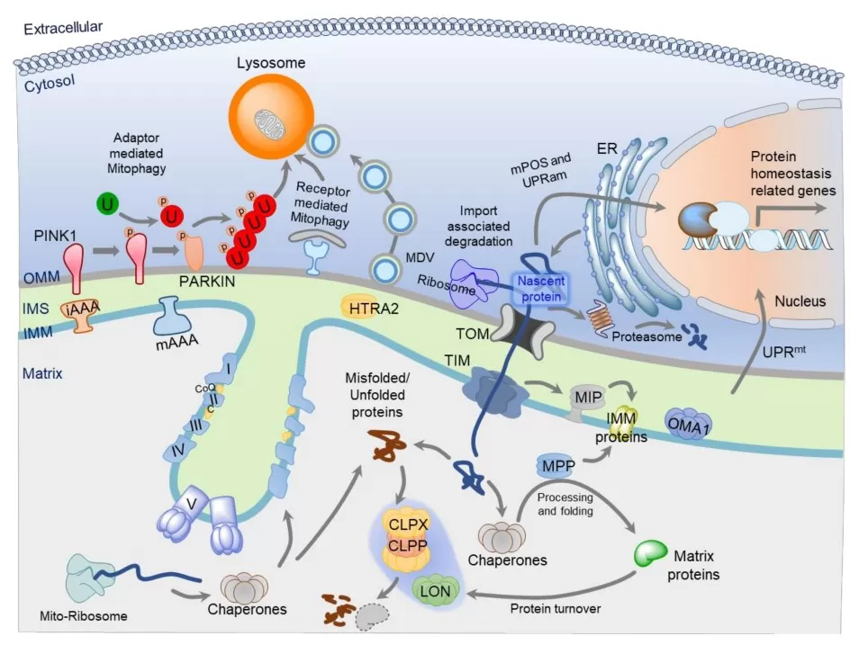
Figure 3. Mitochondrial protein quality-control mechanisms. Import surveillance, folding, proteolysis, stress responses, vesicle transport, and mitophagy act across multiple levels. Reproduced from Figure 1 in Jadiya and Tomar (2020), Genes, 11(5), 563, under CC BY 4.0; resized without content changes.
4. Mitochondrial Proteome Remodeling in Aging and Disease
Aging, neurodegeneration, obesity, and insulin resistance affect several mitochondrial pathways at the same time. The proteins involved and the direction of change vary with tissue composition, cell type, disease stage, and experimental model.
Aging and neurodegeneration. Studies report changes in protein import, mitochondrial translation, respiratory-chain composition, membrane dynamics, oxidative damage, and turnover. The direction and magnitude differ between brain regions and between neuronal and glial cells, so tissue-average proteomics can mask cell-specific defects. Reported changes span protein repair, membrane remodeling, organelle clearance, and mitochondrial replacement (Bogorodskiy et al., 2021; Picca et al., 2023; Picca & Ferrucci, 2026).
Changes in mitochondrial abundance must be separated from changes in composition. A fall in respiratory-chain proteins, for example, may reflect fewer mitochondria, selective loss of one complex, or a shift in neuronal and glial proportions within the sample. Cell-type-resolved or spatial measurements can help distinguish these explanations.
Obesity and insulin resistance. Adipose tissue, liver, and skeletal muscle use different substrates and adapt their mitochondria in different ways. Proteomic studies commonly find changes in OXPHOS, fatty-acid oxidation, TCA-cycle enzymes, redox control, and transport, but the same direction of change can carry a different meaning in each tissue (Li et al., 2020; Picca & Ferrucci, 2026). Mitochondrial content, mtDNA copy number, tissue composition, complex assembly, and functional assays are needed before assigning a change in respiratory capacity or flux (Baker et al., 2024).
Exercise, dietary interventions, and drug treatment can change mitochondrial quantity or substrate preference before broad proteome remodeling becomes visible. Longitudinal sampling, metabolomics, and respiration measurements help distinguish an adaptive response from persistent dysfunction or treatment failure.
5. Mitochondrial Proteomics: Experimental Design and Bioinformatics
The biological question should determine the sample type, enrichment method, acquisition mode, statistical background, and follow-up assays. These choices set both the range of proteins that can be measured and the strength of the conclusions supported by the data.
5.1 Study Design, Enrichment, and LC-MS/MS Quantification
Whole-cell proteomics retains cellular context but may miss low-abundance or compartment-restricted mitochondrial proteins. Differential centrifugation, density gradients, and immunocapture improve mitochondrial coverage, although each method can lose specific proteins, co-isolate other organelles, or disturb membranes and complexes (Nusir et al., 2023). Purity should be checked with positive markers from several mitochondrial compartments and negative markers for the ER, lysosome, nucleus, and cytosol.
Bottom-up workflows generally include protein extraction, reduction and alkylation, digestion, and LC-MS/MS. DDA is widely used for discovery-oriented identification, while DIA offers more complete and reproducible quantification across larger sample sets (Lou & Shui, 2024). Hydrophobic multi-pass proteins, scarce assembly factors, and unstable intermediates remain difficult to detect, so a missing value is not the same as biological absence.
PRM and SRM/MRM provide targeted abundance measurements, Western blotting and immunofluorescence examine selected proteins or localization, and native-complex methods assess assembly state. Respiration, enzyme assays, and metabolite measurements are needed when the claim concerns mitochondrial function or metabolic flux.
Preanalytical handling is especially important after mitochondrial enrichment. Ischemia time, temperature, processing duration, freeze-thaw history, detergent choice, and batch structure can change membrane integrity and recovery. Paired whole-cell and enriched fractions can separate biological abundance changes from altered recovery or redistribution. Reliable comparison also requires biological replication and randomized preparation and injection orders.
5.2 Mitochondrial Annotation and Biological Interpretation
MitoCarta3.0 and MitoPathways provide mitochondrial and sub-organelle annotations. MitoCoP adds copy-number and half-life information, while complexome resources group proteins into import machinery, respiratory assemblies, and assembly intermediates (Rath et al., 2021; Morgenstern et al., 2021; Schulte et al., 2023). GO Cellular Component, interaction networks, and sample-specific evidence provide additional context for these annotations.
Use the proteins that passed detection, quantification, and statistical filtering as the enrichment background. Results are easier to interpret when proteins are grouped into modules such as import, mtDNA maintenance, mitochondrial translation, proteostasis, the TCA cycle, and fatty-acid oxidation. Broad pathway labels alone can hide whether the signal comes from one compartment, one complex, or a general change in mitochondrial content.
Sub-organelle and complex-level annotation can show where a pathway signal is concentrated. A coordinated shift across several subunits is stronger evidence than an isolated protein hit, but even a coherent module must be checked against sample purity, mitochondrial content, and functional data. Claims about OXPHOS capacity, protein-import failure, or mitophagy therefore require evidence beyond coordinated abundance changes.
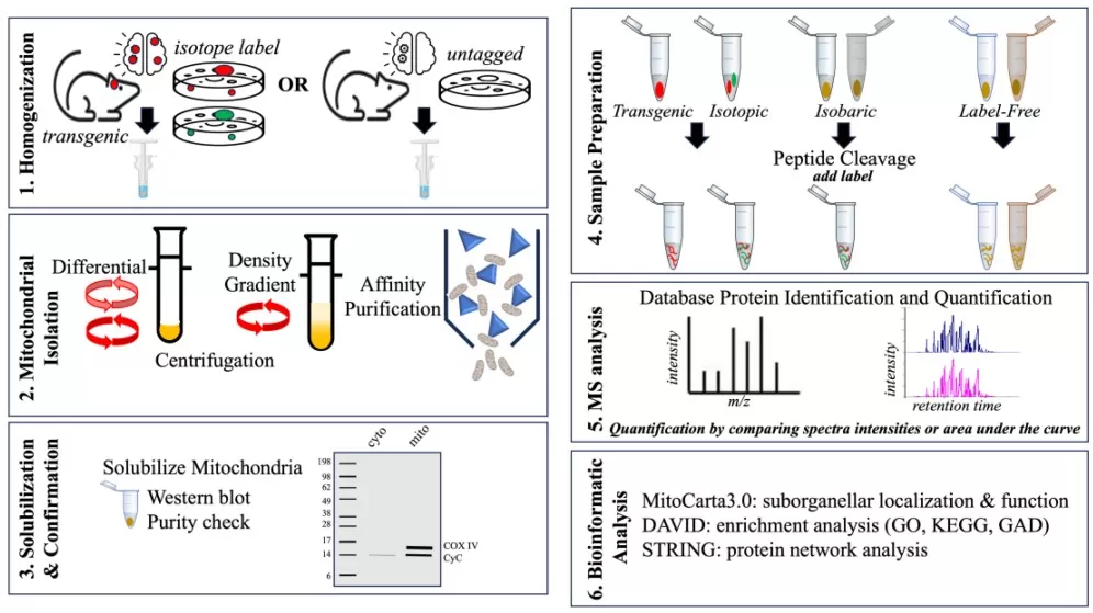
Figure 4. General workflow for mitochondrial proteome analysis. Mitochondrial isolation and purity assessment are followed by quantitative sample preparation, LC-MS analysis, and bioinformatics. Reproduced from Figure 4 in Nusir et al. (2023), Biomolecules, 13(11), 1638, under CC BY 4.0; resized without content changes.
Table 2. Mitochondrial Proteomics Strategies by Research Question
| Research question | Sample and proteomics strategy | Complementary assays | Main interpretation |
|---|---|---|---|
| Mitochondrial composition | Whole-cell or mitochondrial-enriched tissue/cells; DDA or DIA quantitative proteomics | Purity markers; microscopy | Compartment- and function-level protein composition |
| OXPHOS remodeling | Disease-relevant tissue/cells; DIA and complex-focused analysis | Respiration assays; metabolomics | Changes across respiratory complexes and assembly modules |
| Protein-import defects | Cell or tissue models; quantitative proteomics with targeted follow-up | Import assays; membrane potential | Targeting, translocation, and precursor accumulation |
| Mitophagy | Defined perturbation; quantitative and/or ubiquitin/PTM proteomics | Flux reporters; lysosomal inhibition; imaging | Confirm pathway engagement with dynamic flux assays |
| Aging / metabolic disease | Disease-relevant tissues or low-input samples; DIA quantitative proteomics | Transcriptomics; metabolomics; mtDNA; functional assays | Disease-, tissue-, and cell-type-specific remodeling |
How MetwareBio Supports Mitochondrial Proteomics
Before selecting a proteomics workflow, the project should define the sample type, the mitochondrial compartments or protein classes of interest, and the evidence required for the final biological claim.
MetwareBio supports DDA and DIA quantitative proteomics, compatible low-input workflows, and PTM profiling such as phosphoproteomics and acetyl-proteomics. Available downstream analyses include differential-protein analysis, PPI networks, GO/KEGG enrichment, bioinformatics-based subcellular localization, and curated mitochondrial annotation when included in the project scope. Proteomics results may also be integrated with transcriptomics, metabolomics, lipidomics, mtDNA measurements, or functional data.
Studies centered on membrane proteins, respiratory complexes, low-input samples, or PTM regulation require different preparation and validation strategies. Whole-cell and mitochondrial-enriched samples also support different conclusions, and major findings may require targeted or functional follow-up.
Planning a mitochondrial proteomics study? Contact MetwareBio to discuss sample requirements, DDA or DIA quantification, subcellular annotation, PTM profiling, and multi-omics integration.
Contact UsFrequently Asked Questions About Mitochondrial Proteomics
1. How many proteins are present in the human mitochondrial proteome?
There is no single fixed number. MitoCarta3.0 lists 1,136 high-confidence human mitochondrial genes, but tissues and cell states express different subsets and abundances. Quantitative datasets therefore recover overlapping, not identical, mitochondrial protein sets (Rath et al., 2021; Morgenstern et al., 2021).
2. Is mitochondrial proteomics the same as whole-cell proteomics?
No. Whole-cell proteomics keeps the wider cellular context but may underrepresent low-abundance, hydrophobic, or compartment-restricted mitochondrial proteins. Enrichment improves coverage, but it can also introduce selective loss, co-isolation, or damage to membranes and complexes. The better option depends on the question.
3. Can proteomics alone measure mitophagy?
No. Proteomics can detect changes in mitophagy-related proteins, ubiquitination, and pathway modules, but it does not measure flux directly. Dynamic reporters, lysosomal inhibition, microscopy, or other functional assays are needed to determine whether lysosomal delivery and degradation are increased or blocked.
Read More: Proteomics Methods, Quality Control, and Mitochondrial Disease Research
These articles cover complementary topics for researchers planning mitochondrial proteomics projects, from DIA vs DDA method selection and quality control to missing value handling and multi-omics integration in disease studies.
Understand when DIA offers advantages over DDA for large-cohort mitochondrial proteomics, including reproducibility, missing value reduction, and quantitative consistency across multi-batch sample sets.
Learn how combining quantitative proteomics with phosphoproteomics reveals regulatory mechanisms in mitochondrial signaling, OXPHOS modulation, and stress-response pathways beyond abundance alone.
Discover how QC samples, iRT peptides, and batch-monitoring strategies ensure that mitochondrial protein quantification remains reliable across plates, batches, and run sequences.
Explore methods for handling missing values in mitochondrial proteomics datasets, where hydrophobic membrane proteins and low-abundance assembly factors are particularly prone to incomplete detection.
Compare serum and plasma proteomics sample types relevant to mitochondrial biomarker studies, including collection considerations and how matrix choice affects low-abundance protein detection.
See how metabolomics and proteomics reveal mitochondrial metabolic dependence in leukemia, illustrating the integration of mitochondrial proteome analysis with disease mechanism research.
References
- Baker, Z. N., Forny, P., & Pagliarini, D. J. (2024). Mitochondrial proteome research: The road ahead. Nature Reviews Molecular Cell Biology, 25, 65-82. https://doi.org/10.1038/s41580-023-00650-7
- Bogorodskiy, A., Okhrimenko, I., Burkatovskii, D., Jakobs, P., Maslov, I., Gordeliy, V., Dencher, N. A., Gensch, T., Voos, W., Altschmied, J., Haendeler, J., & Borshchevskiy, V. (2021). Role of mitochondrial protein import in age-related neurodegenerative and cardiovascular diseases. Cells, 10(12), 3528. https://doi.org/10.3390/cells10123528
- Giacomello, M., Pyakurel, A., Glytsou, C., & Scorrano, L. (2020). The cell biology of mitochondrial membrane dynamics. Nature Reviews Molecular Cell Biology, 21, 204-224. https://doi.org/10.1038/s41580-020-0210-7
- Jadiya, P., & Tomar, D. (2020). Mitochondrial protein quality control mechanisms. Genes, 11(5), 563. https://doi.org/10.3390/genes11050563
- Lee, H., & Yoon, Y. (2018). Mitochondrial membrane dynamics—Functional positioning of OPA1. Antioxidants, 7(12), 186. https://doi.org/10.3390/antiox7120186
- Li, Y., Ma, Q., Li, P., Wang, J., Wang, M., Fan, Y., Wang, T., Wang, C., Wang, T., & Zhao, B. (2020). Proteomics reveals different pathological processes of adipose tissue, liver, and skeletal muscle under insulin resistance. Journal of Cellular Physiology, 235(10), 6441-6461. https://doi.org/10.1002/jcp.29658
- Lou, R., & Shui, W. (2024). Acquisition and analysis of DIA-based proteomic data: A comprehensive survey in 2023. Molecular & Cellular Proteomics, 23(2), 100712. https://doi.org/10.1016/j.mcpro.2024.100712
- Morgenstern, M., Peikert, C. D., Lübbert, P., et al. (2021). Quantitative high-confidence human mitochondrial proteome and its dynamics in cellular context. Cell Metabolism, 33(12), 2464-2483.e18. https://doi.org/10.1016/j.cmet.2021.11.001
- Nusir, A., Sinclair, P., & Kabbani, N. (2023). Mitochondrial proteomes in neural cells: A systematic review. Biomolecules, 13(11), 1638. https://doi.org/10.3390/biom13111638
- Picca, A., Faitg, J., Auwerx, J., Ferrucci, L., & D'Amico, D. (2023). Mitophagy in human health, ageing and disease. Nature Metabolism, 5, 2047-2061. https://doi.org/10.1038/s42255-023-00930-8
- Picca, A., & Ferrucci, L. (2026). Mitochondrial quality control in human ageing and longevity. Nature Metabolism, 8, 1464-1482. https://doi.org/10.1038/s42255-026-01563-3
- Rath, S., Sharma, R., Calvo, S. E., & Mootha, V. K. (2021). MitoCarta3.0: An updated mitochondrial proteome now with sub-organelle localization and pathway annotations. Nucleic Acids Research, 49(D1), D1541-D1547. https://doi.org/10.1093/nar/gkaa1011
- Schulte, U., den Brave, F., Haupt, A., et al. (2023). Mitochondrial complexome reveals quality-control pathways of protein import. Nature, 614, 153-159. https://doi.org/10.1038/s41586-022-05641-w
- Sim, S. I., Chen, Y., Lynch, D. L., Gumbart, J. C., & Park, E. (2023). Structural basis of mitochondrial protein import by the TIM23 complex. Nature, 621, 620-626. https://doi.org/10.1038/s41586-023-06239-6
- Song, J., Herrmann, J. M., & Becker, T. (2021). Quality control of the mitochondrial proteome. Nature Reviews Molecular Cell Biology, 22, 54-70. https://doi.org/10.1038/s41580-020-00300-2