Uric acid is often used as a routine clinical readout, especially in studies of hyperuricemia and gout. For mechanism-oriented research, however, a single serum uric acid value rarely explains the full biology behind altered urate status. Elevated serum urate can reflect increased purine turnover, xanthine oxidoreductase activity, renal underexcretion, intestinal excretion changes, transporter variation, dietary or drug exposure, or broader metabolic remodeling.
This broader view is important because uric acid sits at the intersection of purine metabolism, kidney function, inflammation, oxidative stress, cardiometabolic disease, and microbiome-associated urate handling. In biomedical and translational studies, uric acid is therefore best interpreted as a context-dependent biomarker rather than an isolated endpoint. This article summarizes uric acid biology, urate homeostasis, disease mechanisms, metabolomics interpretation, LC-MS/MS analysis strategies, and multi-omics research applications.
1. WHAT IS URIC ACID? CHEMICAL FEATURES AND PURINE METABOLISM
Uric acid is generated during the breakdown of purine nucleotides. AMP- and GMP-derived intermediates are converted through nucleosides and purine bases, leading to hypoxanthine and xanthine. Xanthine oxidoreductase catalyzes the final two oxidative steps, converting hypoxanthine to xanthine and xanthine to uric acid. In humans and other hominids, uricase is inactive, so uric acid is not further degraded to allantoin as efficiently as in many other mammals (Wen et al., 2024; Liu et al., 2023b).
Because purine degradation is linked to nucleotide turnover, energy balance, cell injury, diet, and renal excretion, uric acid can respond to multiple biological inputs. This explains why uric acid is relevant not only to gout research, but also to studies of kidney disease, metabolic stress, inflammation, and pharmacodynamic responses to xanthine oxidase inhibitors.
Uric acid and urate are closely related terms, but their chemical context matters. At physiological pH, uric acid exists largely as urate, whereas low-pH environments favor the less soluble protonated form. This acid-base behavior is important for understanding uric acid solubility, crystallization risk, renal handling, and uric acid stone formation (Halperin Kuhns & Woodward, 2021; Dalbeth et al., 2021).
These chemical and pathway features set up a central research question: whether altered urate status mainly reflects purine production, transport, excretion, or disease-associated metabolic remodeling. The following section therefore examines uric acid homeostasis from a systems-level perspective.
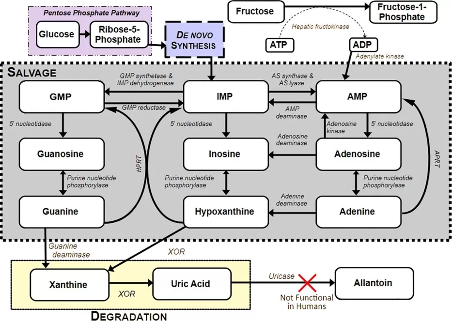
Figure 1. Overview of purine metabolism and uric acid production. Image reproduced from Nelson and Voruganti, 2026, Frontiers in Endocrinology, 16, 1662037, licensed under the Creative Commons Attribution License (CC BY).
2. URIC ACID HOMEOSTASIS: PURINE PRODUCTION, URATE TRANSPORT, AND EXCRETION
Uric acid homeostasis is controlled by production, renal excretion, intestinal excretion, and transporter activity. This systems-level framing is more informative than describing uric acid only as a metabolic waste product because the same change in serum urate can have different mechanistic origins.
2.1 Purine Degradation and Xanthine Oxidoreductase in Uric Acid Production
Purine degradation provides the biochemical source of uric acid. Nucleotide turnover generates inosine, hypoxanthine, xanthine, and other intermediates, while xanthine oxidoreductase completes uric acid formation. In oxidative stress and metabolic stress contexts, this enzyme system is frequently discussed because xanthine oxidase activity can be associated with reactive oxygen species generation. However, causality should be phrased carefully; uric acid, xanthine oxidoreductase activity, and oxidative stress may be connected differently depending on tissue, disease state, and experimental model (Wen et al., 2024; Du et al., 2024).
2.2 Renal and Intestinal Urate Handling: Transporters and the Gut-Kidney Axis
The kidney is central to serum urate regulation. Filtration, reabsorption, and secretion in renal tubules shape circulating urate levels, and urate transporters such as URAT1/SLC22A12, GLUT9/SLC2A9, and ABCG2 are important determinants of urate handling. Genetic and pharmacological studies have made these transporters important targets for understanding hyperuricemia phenotypes and drug response (Halperin Kuhns & Woodward, 2021; Leask et al., 2024).
The intestine also contributes to extra-renal urate elimination. Recent microbiome research has shown that gut bacterial pathways can degrade uric acid and may partially compensate for uricase loss in hominids, supporting interest in the gut-kidney axis in hyperuricemia and gout studies (Liu et al., 2023b).
Table 1. Biological and Analytical Determinants of Serum Uric Acid Levels
| Determinant | Related Process | Example Factors / Molecules | Research Meaning |
|---|---|---|---|
| Purine production | Nucleotide turnover and degradation | Hypoxanthine, xanthine, xanthine oxidoreductase | Helps evaluate uric acid overproduction and purine pathway activity |
| Renal excretion | Filtration, secretion, and reabsorption | URAT1, GLUT9, OAT family, renal function | Helps distinguish renal underexcretion phenotypes |
| Intestinal excretion | Extra-renal urate elimination | ABCG2, gut-kidney axis, microbiome-related factors | Supports interpretation of non-renal urate handling |
| Diet and exposure | Purine/fructose intake and drug effects | Dietary purines, fructose, xanthine oxidase inhibitors | Important for study design and confounder control |
| Disease context | Metabolic and inflammatory status | CKD, obesity, diabetes, inflammation | Places serum uric acid in broader disease mechanisms |
3. URIC ACID IN DISEASE MECHANISMS: HYPERURICEMIA, GOUT, KIDNEY STONES, AND CARDIOMETABOLIC RESEARCH
Uric acid is relevant to disease research because it links biochemical metabolism with tissue-specific pathology. Rather than providing a disease catalogue, this section focuses on how uric acid-related readouts can support mechanism studies, biomarker discovery, urinary metabolome research, and intervention analysis.
3.1 Hyperuricemia and Gout: From Urate Crystal Deposition to Biomarker Discovery
Hyperuricemia is the key biochemical risk factor for gout, but high serum urate alone does not mean that gout will occur. Gout develops through a sequence that includes sustained hyperuricemia, monosodium urate crystal deposition, and crystal-driven innate immune activation. MSU crystals can activate inflammatory pathways that include the NLRP3 inflammasome and IL-1beta signaling, which are central to gout flare biology (Dalbeth et al., 2021; Leask et al., 2024).
3.2 Kidney Disease, Uric Acid Stones, and Urinary Metabolome Research
The kidney is both a regulator and a target of uric acid biology. In uric acid stone research, urine pH, urine dilution, ammonium handling, organic acid background, and local renal pelvis chemistry may be more informative than serum uric acid alone. A 2024 paired-sample multi-omics study of renal pelvis urine from patients with unilateral uric acid stones used LC-MS/MS-based proteomics and metabolomics to characterize a local urinary microenvironment associated with stone formation (Xu et al., 2024).
This type of research highlights the value of serum and urine as complementary matrices. Serum or plasma can reflect systemic urate burden and metabolic exposure, while urine provides information on renal excretion phenotype, acid-base status, dilution effects, and stone-associated microenvironments.
3.3 Cardiometabolic Disease, Metabolic Syndrome, and Nutritional Exposure
Serum uric acid has been studied in relation to obesity, insulin resistance, metabolic syndrome, hypertension, chronic kidney disease, diabetes, and cardiovascular disease. These associations should be written with careful language because observational relationships do not always establish causality. In research design, uric acid can be interpreted alongside lipid metabolism, amino acid metabolism, energy metabolism, renal function, and inflammatory markers to understand disease-associated metabolic remodeling (Du et al., 2024; Wen et al., 2024).
Dietary purines, fructose exposure, alcohol intake, renal function, and medication use should be controlled or recorded when possible. Without these variables, pathway-level interpretation can be confounded, especially in studies of cardiometabolic disease and nutritional exposure.
3.4 Drug Response and Uric Acid-Lowering Intervention Research
Uric acid-lowering interventions create a clear application space for targeted metabolomics. Xanthine oxidase inhibitors and other urate-lowering strategies can alter uric acid together with upstream metabolites such as hypoxanthine, xanthine, inosine, and adenosine. Monitoring a purine pathway panel before and after intervention can support mechanism validation, pharmacodynamic assessment, and response heterogeneity analysis. In research settings, these profiles support mechanistic and pharmacodynamic interpretation rather than replacing clinical decision-making.
3.5 Uric Acid as a Context-Dependent Biomarker
Uric acid has context-dependent biology. In plasma, it can contribute to antioxidant capacity; in other settings, elevated urate, crystal deposition, and xanthine oxidoreductase activity may be associated with oxidative stress, inflammation, and tissue pathology. Interpretation should therefore consider sample type, disease stage, renal function, diet, drug exposure, and related pathway metabolites (Wen et al., 2024; Du et al., 2024).
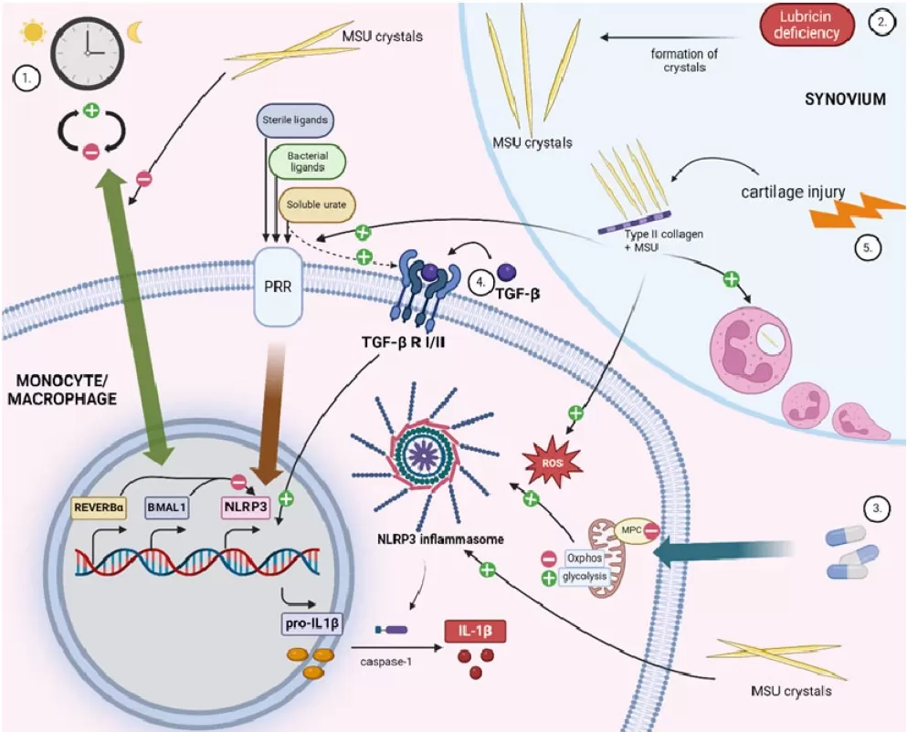
Figure 2. Highlights of inflammatory mechanisms in gout. Image reproduced from Muntiu et al., 2024, Gout, Urate, and Crystal Deposition Disease, 2(3), 220-235, licensed under the Creative Commons Attribution 4.0 International License (CC BY 4.0). Figure created with BioRender.com in the original article.
4. BIOLOGICAL INTERPRETATION OF URIC ACID METABOLOMICS: FROM SINGLE READOUT TO MECHANISTIC CONTEXT
This section focuses on how uric acid-related metabolomics data should be interpreted biologically. The key question is not which instrument is used, but what a measured change in uric acid means in relation to purine turnover, renal handling, disease context, and related molecular layers. Analytical platforms and quality-control decisions are addressed separately in Section 5.
4.1 Purine Pathway Metabolites Beyond Uric Acid
Uric acid becomes more informative when interpreted together with upstream purine pathway metabolites. Hypoxanthine, xanthine, inosine, adenosine, and AMP- or GMP-related metabolites can help distinguish increased nucleotide turnover, altered purine salvage, energy stress, or xanthine oxidoreductase inhibition. This pathway-level interpretation is especially useful when serum uric acid alone cannot explain whether a phenotype is driven mainly by production, excretion, or intervention response (Li et al., 2024).
4.2 How Sample Choice Shapes Uric Acid Interpretation and Study Design
Because uric acid biology is compartment-dependent, sample choice should be treated as part of study design rather than as a late technical decision. Serum and plasma are useful for evaluating systemic urate burden, biomarker discovery, and cardiometabolic associations. Urine is more directly linked to renal excretion, urine chemistry, acid-base status, and stone-forming microenvironments. Tissue, cell, and synovial fluid samples can support more local mechanistic questions, including transporter regulation, enzyme activity, crystal-driven inflammation, and drug response. Matching the sample matrix to the research question helps prevent a serum-centered interpretation from being applied to mechanisms that are primarily renal, urinary, or tissue-localized.
4.3 Multi-Omics Integration for Mechanism-Level Interpretation
Multi-omics integration is most useful when metabolite changes require mechanistic explanation. Metabolomics can define changes in purine metabolism, energy metabolism, amino acid metabolism, lipid metabolism, and microbiome-associated metabolites. Transcriptomics and proteomics can clarify whether enzymes, urate transporters, renal tubular pathways, or inflammatory proteins are altered. Microbiome data can add information about intestinal urate degradation and the gut-kidney axis. In hyperuricemia research, integrated serum and urine metabolomics with transcriptomic data has been used to construct regulatory networks and identify candidate mechanisms (Liu et al., 2023a).
Table 2. Interpretation Framework for Uric Acid Metabolomics and Multi-Omics Research
| Research Question | Biological Layer | Key Readouts | Interpretation Focus |
|---|---|---|---|
| Hyperuricemia mechanism | Systemic urate + renal handling | Serum/urine uric acid; hypoxanthine; xanthine; renal function | Production vs. excretion |
| Gout inflammation | Crystal-driven inflammation | MSU crystal context; urate; IL-1beta/NLRP3-related proteins | Innate immune activation |
| Uric acid stones | Urinary microenvironment | Urinary uric acid; urine pH; organic acids; proteins/metabolites | Stone-forming environment |
| Drug response | Purine pathway modulation | Uric acid; xanthine; hypoxanthine; inosine; adenosine | XOR inhibition or urate-lowering response |
| Cardiometabolic context | Metabolic remodeling | Uric acid; lipid, amino acid, energy, and inflammatory markers | Broader metabolic phenotype |
5. ANALYTICAL WORKFLOW FOR URIC ACID AND PURINE METABOLITE PROFILING: PLATFORM SELECTION, QUANTIFICATION, AND QUALITY CONTROL
After the biological question has been defined, the next step is to choose an analytical workflow that can answer it reliably. Section 5 therefore focuses on measurement strategy: clinical chemistry versus LC-MS/MS, targeted versus discovery-oriented metabolomics, sample handling, normalization, and quality control. This workflow-oriented perspective complements the biological interpretation framework above by focusing on how reliable uric acid and purine metabolite data are generated.
5.1 Clinical Chemistry Assays vs Research-Grade LC-MS/MS
Clinical chemistry assays are efficient for high-throughput serum uric acid measurement and remain important in clinical settings. Research-grade LC-MS/MS provides higher molecular specificity, the ability to measure upstream and downstream metabolites together, isotope-labeled internal standard correction, and stronger compatibility with pathway interpretation. Platform choice should therefore depend on whether the goal is routine uric acid monitoring, mechanistic purine metabolism analysis, biomarker discovery, or intervention response profiling.
5.2 Targeted LC-MS/MS for Absolute Quantification of Uric Acid and Purine Metabolites
Targeted LC-MS/MS workflows commonly use triple quadrupole instruments, multiple reaction monitoring, chemical standards, calibration curves, stable-isotope internal standards, and quality control samples. Hydrophilic interaction chromatography can be useful for separating polar purine metabolites, and validated targeted methods have been developed for simultaneous quantification of multiple purine metabolites in plasma and urine (Li et al., 2024). This approach is most appropriate when predefined analytes require sensitive, reproducible, and quantitative measurement.
5.3 Untargeted and Widely Targeted Metabolomics for Discovery-Oriented Studies
Untargeted metabolomics is appropriate when the research question extends beyond uric acid and the purine pathway. It can reveal broader metabolic shifts in amino acid metabolism, lipid metabolism, central carbon metabolism, oxidative stress-related pathways, and microbiome-associated metabolites. Widely targeted metabolomics can provide broader metabolite coverage with improved quantitative consistency compared with purely untargeted feature discovery, making it suitable for translational studies that require both coverage and reproducibility. These discovery-oriented workflows are most useful when the study requires broader metabolic coverage while maintaining an analytical design suitable for downstream biological interpretation.
5.4 Pre-Analytical Variables, Normalization, and Quality Control
Reliable uric acid and purine metabolite analysis requires attention to fasting status, dietary purine and fructose exposure, renal function, medication use, hemolysis, sample processing time, freeze-thaw history, batch effects, and matrix-specific normalization. Urine studies require additional attention to dilution, pH, creatinine normalization, specific gravity, and collection timing. These factors should be documented before statistical modeling and biological interpretation, because uncontrolled pre-analytical variation can be mistaken for true biological differences.
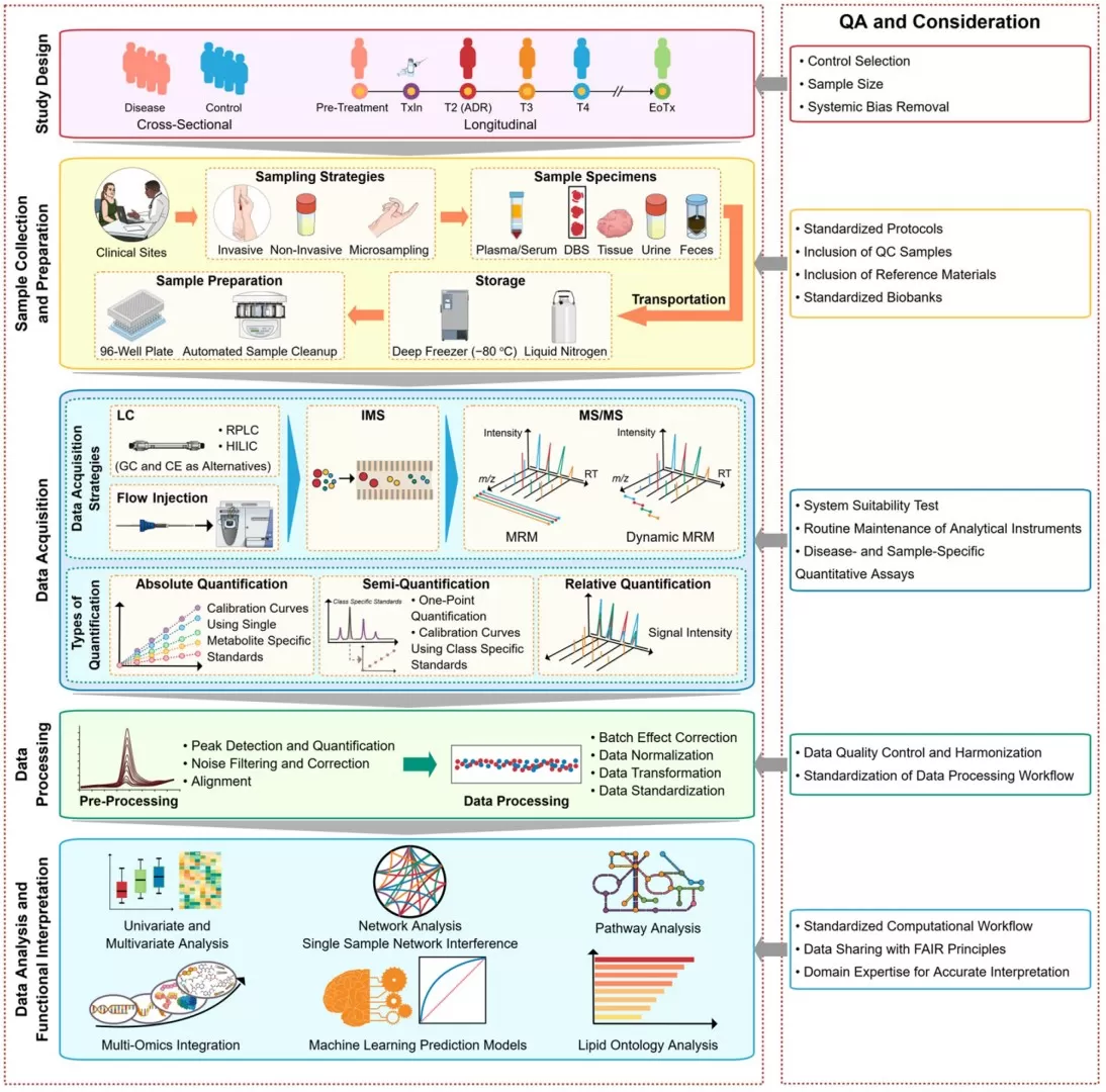
Figure 3. Suggested framework for high-throughput clinical targeted metabolomics and lipidomics studies. The workflow covers study design, biological sample collection and preparation, LC-MS/MS data acquisition, quantitative strategies, data processing, functional interpretation, and major quality assurance considerations. Reproduced from Anh et al. (2024), licensed under the Creative Commons Attribution 4.0 International License (CC BY 4.0).
How MetwareBio Supports Uric Acid, Purine Metabolism, and Multi-Omics Research
Uric acid research often requires more than a single concentration measurement. Mechanism-oriented projects may need to connect uric acid with purine metabolism, renal handling, energy metabolism, inflammation, microbiome-related urate degradation, and disease-associated metabolic remodeling. MetwareBio's metabolomics and multi-omics services can support these goals through LC-MS-based metabolomics, targeted metabolomics, untargeted metabolomics, widely targeted metabolomics, proteomics, transcriptomics, and integrated multi-omics analysis.
For projects focused on uric acid or purine-related metabolites, analytical planning should begin with the research question, sample type, target metabolite coverage, quantification strategy, and quality control design. Serum and urine paired analysis can be considered when production and excretion mechanisms are both relevant. For gout, kidney stone, cardiometabolic, or intervention-response studies, metabolomics can be combined with proteomics, transcriptomics, microbiome profiling, or genomics to strengthen biological interpretation.
Interested in uric acid, purine metabolism, or hyperuricemia-related metabolomics research? Contact MetwareBio to discuss sample type, analytical strategy, targeted metabolite coverage, quality control design, and multi-omics integration options for a research project.
Contact UsFAQ: Uric Acid Metabolism, Hyperuricemia, and Multi-Omics Research
What is uric acid metabolism?
Uric acid metabolism refers to the biochemical process by which purine nucleotides are degraded into uric acid. AMP- and GMP-derived metabolites are converted through intermediates such as inosine, hypoxanthine, and xanthine before xanthine oxidoreductase catalyzes the final steps of uric acid production. In research, this pathway is important for studying purine turnover, oxidative stress, renal handling, and metabolic disease mechanisms.
What is the difference between uric acid and urate?
Uric acid and urate refer to closely related chemical forms. At physiological pH, most uric acid exists as urate, its deprotonated and more soluble form. In lower-pH environments, the protonated uric acid form becomes more relevant and less soluble. This distinction matters for understanding urate transport, crystallization risk, kidney stone formation, and sample-specific interpretation.
Why does serum uric acid increase in hyperuricemia?
Serum uric acid can increase when uric acid production is elevated, excretion is reduced, or both processes occur together. Mechanisms may include increased purine turnover, higher xanthine oxidoreductase activity, renal underexcretion, altered intestinal urate elimination, transporter variation, diet, medication exposure, or broader metabolic remodeling. Mechanism-oriented studies often require related purine metabolites, renal markers, and sample context.
How does uric acid contribute to gout inflammation?
Gout develops when sustained hyperuricemia promotes monosodium urate crystal deposition in joints or tissues. These crystals can activate innate immune responses, including NLRP3 inflammasome signaling and IL-1β production, leading to inflammatory flares. However, elevated serum urate alone does not fully explain gout biology, so inflammatory markers, crystal context, disease stage, and tissue-specific responses should also be considered.
Why are serum and urine both useful in uric acid research?
Serum and urine provide complementary information in uric acid research. Serum or plasma can reflect systemic urate burden, metabolic exposure, and biomarker associations. Urine is more directly related to renal excretion, dilution, urine pH, acid-base status, and stone-forming microenvironments. Paired serum and urine analysis can help distinguish production-driven and excretion-related mechanisms.
How can LC-MS/MS be used to study uric acid and purine metabolites?
LC-MS/MS can quantify uric acid together with related purine metabolites such as hypoxanthine, xanthine, inosine, and adenosine. Compared with a single uric acid measurement, targeted LC-MS/MS provides stronger pathway-level information for studying purine degradation, xanthine oxidoreductase-related changes, intervention response, and sample-specific metabolic profiles. Proper calibration, internal standards, and quality control are essential for reliable interpretation.
Why is multi-omics useful for hyperuricemia, gout, and uric acid stone research?
Multi-omics can connect uric acid changes with broader biological mechanisms. Metabolomics can profile purine metabolism, energy metabolism, inflammation-related metabolites, and urinary microenvironment changes. Proteomics and transcriptomics can reveal changes in urate transporters, inflammatory proteins, renal pathways, or enzyme regulation. Microbiome analysis may add information about intestinal urate degradation and the gut-kidney axis.
Read More: Metabolomics and Multi-Omics for Metabolic Biomarker Research
The following articles extend the analytical and biological themes discussed above, covering platform selection, metabolomics strategy comparison, kidney biomarker research, gut-kidney axis profiling, and multi-omics integration for metabolic disease studies.
Section 5 of this article discusses targeted LC-MS/MS, untargeted metabolomics, and widely targeted metabolomics as complementary analytical strategies. This guide compares all three approaches in depth, covering coverage, sensitivity, quantification accuracy, and suitability for different research questions.
Uric acid and purine metabolite profiling relies heavily on LC-MS/MS, but understanding when GC-MS is more appropriate helps researchers design better analytical workflows. This comparison covers ionization methods, analyte compatibility, and platform selection criteria for metabolomics studies.
Like uric acid, creatinine is a clinically important metabolite linked to kidney function. This article explores creatinine's role as a renal biomarker, its relationship with muscle metabolism, and how metabolomics can contextualize creatinine alongside other nitrogen metabolism readouts.
The gut-kidney axis discussed in Section 2.2 highlights how intestinal microbiome pathways contribute to urate degradation. This service overview explains how combined microbiome sequencing and metabolomics profiling can reveal gut-derived metabolite contributions to systemic metabolic phenotypes.
Section 4.3 describes how multi-omics integration strengthens mechanism-level interpretation of uric acid data. This article provides a detailed framework for combining proteomics and metabolomics, covering correlation analysis, pathway mapping, and biological context building.
For uric acid research spanning hyperuricemia, gout, kidney stones, and cardiometabolic disease, multi-omics approaches connect metabolite changes with protein and transcript-level regulation. This article reviews the technological advantages and application areas of integrated multi-omics analysis across disease research.
References
- Dalbeth, N., Gosling, A. L., Gaffo, A., & Abhishek, A. (2021). Gout. The Lancet, 397(10287), 1843–1855. https://doi.org/10.1016/S0140-6736(21)00569-9
- Du, L., Yao, Z., Li, H., Wang, Q., Xie, L., Yang, B., Pang, Y., Zhang, C., Zhong, Z., & Gao, J. (2024). Hyperuricemia and its related diseases: mechanisms and advances in therapy. Signal Transduction and Targeted Therapy, 9, 212. https://doi.org/10.1038/s41392-024-01916-y
- Halperin Kuhns, V. L., & Woodward, O. M. (2021). Urate transport in health and disease. Best Practice & Research Clinical Rheumatology, 35(4), 101717. https://doi.org/10.1016/j.berh.2021.101717
- Leask, M. P., Crisan, T. O., Ji, A., Matsuo, H., Kottgen, A., & Merriman, T. R. (2024). The pathogenesis of gout: molecular insights from genetic, epigenomic and transcriptomic studies. Nature Reviews Rheumatology, 20, 510-523. https://doi.org/10.1038/s41584-024-01137-1
- Li, X., Liu, Z., Li, Z., Xiong, X., Zhang, X., Yang, C., Zhao, L., & Zhao, R. (2024). A simple, rapid and sensitive HILIC LC-MS/MS method for simultaneous determination of 16 purine metabolites in plasma and urine. Talanta, 267, 125171. https://doi.org/10.1016/j.talanta.2023.125171
- Liu, H., Xie, R., Dai, Q., Fang, J., Xu, Y., & Li, B. (2023a). Exploring the mechanism underlying hyperuricemia using comprehensive research on multi-omics. Scientific Reports, 13, 7161. https://doi.org/10.1038/s41598-023-34426-y
- Liu, Y., Jarman, J. B., Low, Y. S., Augustijn, H. E., Huang, S., Chen, H., & Dodd, D. (2023b). A widely distributed gene cluster compensates for uricase loss in hominids. Cell, 186, 3400-3413.e20. https://doi.org/10.1016/j.cell.2023.06.010
- Wen, S., Arakawa, H., & Tamai, I. (2024). Uric acid in health and disease: From physiological functions to pathogenic mechanisms. Pharmacology & Therapeutics, 256, 108615. https://doi.org/10.1016/j.pharmthera.2024.108615
- Xu, S., Liu, Z.-L., Zhang, T.-W., Li, B., Wang, X.-N., & Jiao, W. (2024). Self-control study of multi-omics in identification of microenvironment characteristics in urine of uric acid stone. Scientific Reports, 14, 25165. https://doi.org/10.1038/s41598-024-76054-0
- Nelson, K. L., & Voruganti, V. S. (2026). Implication of xanthine oxidoreductase in oxidative stress-related chronic diseases. Frontiers in Endocrinology, 16, 1662037. https://doi.org/10.3389/fendo.2025.1662037
- Muntiu, M., Joosten, L. A. B., & Crisan, T. O. (2024). Gout basic research: 2023 in review. Gout, Urate, and Crystal Deposition Disease, 2(3), 220-235. https://doi.org/10.3390/gucdd2030017
- Anh, N. K., Thu, N. Q., Tien, N. T. N., Long, N. P., & Nguyen, H. T. (2024). Advancements in mass spectrometry-based targeted metabolomics and lipidomics: Implications for clinical research. Molecules, 29(24), 5934. https://doi.org/10.3390/molecules29245934