Oxidative stress cannot be characterized by a single reactive oxygen species (ROS) measurement. Reduced glutathione (GSH) and oxidized glutathione (GSSG) form an interconnected redox couple reflecting peroxide detoxification, thiol chemistry, and reducing capacity. Because GSH may oxidize after collection and GSSG is typically much less abundant, sample handling can substantially alter the apparent balance. Reliable interpretation therefore requires biochemical context, rapid stabilization, and an analytical strategy matched to the biological question.
1. Reduced vs. Oxidized Glutathione: Structure and Key Differences
GSH and GSSG differ in oxidation state and chemical reactivity but remain interconvertible components of the same glutathione system.
Reduced glutathione (GSH). Reduced glutathione is a tripeptide composed of glutamate, cysteine, and glycine, with glutamate and cysteine joined through a γ-glutamyl linkage. Its cysteinyl thiol is the key reactive center, supporting reduction reactions, electrophile conjugation, and thiol-disulfide exchange. GSH serves as a substrate for glutathione peroxidases and glutathione S-transferases and participates in reversible protein S-glutathionylation (Lu, 2013).
Oxidized glutathione (GSSG). GSSG forms when two GSH molecules are linked by a disulfide bond between their cysteine residues. It is produced during normal glutathione-dependent peroxide reduction and can be converted back to GSH by glutathione reductase. GSSG is therefore a functional intermediate in redox cycling rather than a metabolic waste product.
GSSG accumulation shifts the glutathione pool toward a more oxidized state, although its interpretation remains context dependent.
Table 1. Key Biochemical Differences Between GSH and GSSG
| Feature | GSH | GSSG |
|---|---|---|
| Full name | Reduced glutathione | Oxidized glutathione, or glutathione disulfide |
| Redox state | Reduced | Oxidized |
| Structural feature | One glutathione tripeptide with a free cysteinyl thiol | Two glutathione molecules linked by a cysteine-cysteine disulfide bond |
| Main reactive group | Thiol (-SH) | Disulfide (-S-S-) |
| Major biological role | Reducing substrate, peroxide detoxification, electrophile conjugation, protein-thiol regulation | Intermediate in glutathione redox cycling and indicator of oxidation within the glutathione pool |
| Relative abundance | Usually the predominant intracellular form | Usually much less abundant than GSH |
| Analytical behavior | Susceptible to ex vivo oxidation during collection and processing | Low abundance and vulnerable to artifactual elevation when GSH oxidizes after sampling |
| Research interpretation | Reflects pool size, synthesis, consumption, conjugation, export, and adaptive regulation | Reflects oxidation-reduction balance, regeneration capacity, export, and preanalytical handling |
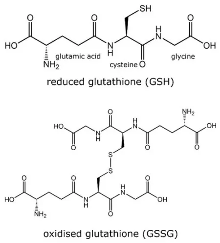
Figure 1. Chemical structures of reduced glutathione (GSH) and oxidized glutathione (GSSG). GSH consists of γ-glutamyl, cysteinyl, and glycine residues, whereas GSSG contains two glutathione molecules linked by a disulfide bond between their cysteine residues. Reproduced from Al-Temimi et al. (2023), Metabolites, 13(4), 465, under the Creative Commons Attribution 4.0 International License (CC BY 4.0).
2. GSH-GSSG Metabolism and Redox Homeostasis
The glutathione pool is determined by more than synthesis alone. Its state reflects coordinated synthesis, oxidation, reduction, conjugation, export, compartmental transport, and NADPH regeneration. Consequently, a change in GSH or GSSG can arise from altered substrate availability or enzyme activity even when ROS production is unchanged.
2.1 GSH Biosynthesis and Cysteine Supply
GSH synthesis proceeds through two ATP-dependent cytosolic reactions. Glutamate-cysteine ligase (GCL) forms γ-glutamylcysteine from glutamate and cysteine, after which glutathione synthetase adds glycine. Cysteine availability and GCL activity are major control points (Lu, 2013).
Cysteine may be obtained from extracellular cysteine or cystine and, in some tissues, from the methionine cycle through the transsulfuration pathway. The SLC7A11-containing cystine/glutamate antiporter system xc− supports cysteine availability after imported cystine is reduced, although its contribution depends on nutrient conditions and transporter expression (Jyotsana et al., 2022).
2.2 GSH Oxidation in GPX-Dependent Peroxide Detoxification
Glutathione peroxidases use GSH to reduce hydrogen peroxide or organic hydroperoxides, oxidizing two GSH molecules to form one GSSG molecule. GPX4 extends this chemistry to phospholipid hydroperoxides and helps limit iron-dependent lipid peroxidation and ferroptosis (Jiang et al., 2021).
GSH is also consumed through glutathione S-transferase-catalyzed conjugation and reversible protein S-glutathionylation. Low free GSH may therefore reflect conjugation, protein binding, degradation, or export rather than conversion to GSSG alone.
2.3 NADPH-Dependent GSSG Reduction and GSH Regeneration
Glutathione reductase converts GSSG back to GSH using NADPH. Redox balance therefore depends on cellular NADPH-generating pathways, with the oxidative pentose phosphate pathway often making a major contribution and other pathways contributing in a cell- and context-dependent manner.
A lower GSH/GSSG ratio may reflect increased peroxide burden, impaired GSSG reduction, inadequate NADPH regeneration, limited cysteine supply, or several changes acting together. It is therefore an integrated metabolic readout rather than a direct ROS meter.
2.4 Compartment-Specific GSH/GSSG Pools
Glutathione pools differ across the cytosol, mitochondria, nucleus, endoplasmic reticulum, and extracellular space; whole-cell or tissue-homogenate measurements may therefore obscure compartment-specific redox changes.
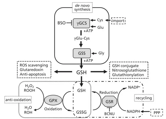
Figure 2. Roles and metabolism of glutathione, including de novo synthesis, GPX-dependent peroxide reduction, GSSG formation, glutathione reductase-dependent recycling, and pentose phosphate pathway-supported NADPH regeneration. Reproduced from Fujii et al. (2011), Journal of Clinical Biochemistry and Nutrition, 49(2), 70-78, under the Creative Commons Attribution License.
Abbreviations: BSO, buthionine sulfoximine; BCNU, carmustine; γGCS, γ-glutamylcysteine synthetase; GSS, glutathione synthetase; GPX, glutathione peroxidase; GSR, glutathione reductase; PPP, pentose phosphate pathway.
3. Interpreting GSH, GSSG, and the GSH/GSSG Ratio
The selected readout determines what can be concluded from a glutathione experiment. GSH, GSSG, total glutathione, the GSH/GSSG ratio, and glutathione redox potential are related but not interchangeable. Each captures a different aspect of pool size, oxidation, or thermodynamic state.
3.1 Biological Interpretation of GSH and GSSG Concentrations
A decrease in GSH can result from reduced synthesis, oxidation, conjugation, protein S-glutathionylation, export, degradation, or loss of cellular integrity. Increased GSH may reflect adaptive synthesis. Absolute concentration should therefore be interpreted alongside GSSG, total pool size, sample viability, and pathway context.
Higher GSSG is often consistent with oxidation, but GSSG is highly sensitive to preanalytical error. Delayed processing or inadequate thiol stabilization can generate GSSG after collection, so interpretation requires standardized handling and adequate analytical performance at the lower end of the GSSG range (Tomin et al., 2020; Thiel et al., 2023).
3.2 GSH/GSSG Ratio vs. Glutathione Redox Potential
The molar GSH/GSSG ratio is a common relative indicator of redox balance. A lower ratio generally indicates a more oxidized glutathione pool, but no universal reference interval applies across species, tissues, cells, or biofluids. Comparisons are strongest within a controlled study using one matrix, protocol, analytical platform, and normalization method.
The ratio is not identical to the redox potential of the 2GSH/GSSG couple. Redox potential is calculated from the Nernst relationship and depends on the square of GSH concentration, GSSG concentration, temperature, and pH. Two samples can therefore have the same GSH/GSSG ratio but different redox potentials if their absolute pool sizes differ (Jones, 2002). For many metabolomics studies, the ratio is a practical comparative readout, whereas redox potential provides a more explicit thermodynamic description when concentration and pH data are sufficiently reliable.
Table 2. Interpretation and Limitations of Common Glutathione Redox Readouts
| Readout | What it primarily reflects | Main limitation | Recommended interpretation |
|---|---|---|---|
| GSH concentration | Size of the reduced glutathione pool | Influenced by synthesis, consumption, conjugation, export, cell number, and sample oxidation | Interpret with GSSG, total glutathione, viability, and pathway context |
| GSSG concentration | Oxidized component of the glutathione pool | Low abundance and highly sensitive to ex vivo GSH oxidation | Use only with validated stabilization and adequate lower-range analytical performance |
| Total glutathione | Overall pool size; often expressed as GSH + 2 × GSSG in GSH equivalents | Calculation and units can vary by method | State the calculation convention and use it to distinguish pool depletion from redistribution |
| GSH/GSSG ratio | Relative distribution between reduced and oxidized forms | Does not incorporate pH or absolute pool size and has no universal normal range | Use for controlled within-study comparisons |
| Glutathione redox potential | Thermodynamic state of the 2GSH/GSSG couple | Requires accurate concentrations, pH, temperature, and equilibrium assumptions | Use when the study is designed for quantitative redox thermodynamics |
4. GSH/GSSG Balance in Disease and Cellular Stress
Altered glutathione metabolism is observed in many disease models, but its direction and biological significance are context dependent. A change in GSH/GSSG can reflect tissue injury, metabolic limitation, compensatory defense, treatment response, or a combination of these processes. Disease interpretation should therefore connect the glutathione readout to mechanism and phenotype rather than treating it as a stand-alone diagnostic marker.
4.1 GSH/GSSG Regulation in Cancer, Treatment Resistance, and Ferroptosis
Some cancer cells increase SLC7A11-dependent cystine uptake, GSH synthesis, NADPH production, or glutathione peroxidase activity under oxidative pressure. These adaptations can support survival and resistance to selected treatments, with effects varying by cell lineage, genotype, nutrient availability, and drug mechanism (Jyotsana et al., 2022).
The GSH-GPX4 axis is closely linked to ferroptosis. Restricting cysteine, depleting GSH, or inhibiting GPX4 can increase phospholipid peroxidation in responsive cells, but sensitivity also depends on lipid composition, iron metabolism, parallel antioxidant systems, and tissue context (Jiang et al., 2021).
4.2 GSH/GSSG Imbalance in Aging and Neurodegenerative Disease
Because the brain has high energy demand, abundant oxidizable lipids, and strong dependence on mitochondrial function, glutathione metabolism is frequently studied in aging and neurodegeneration. Regional and cell-type differences are substantial, and observed changes may involve synthesis, transport, mitochondrial balance, or neuroinflammation (Aoyama, 2021).
Peripheral-blood GSH or GSSG is not a direct proxy for the central nervous system. Brain-focused conclusions require an appropriate combination of neural tissue, cerebrospinal fluid, imaging, or mechanistic models.
4.3 GSH/GSSG Balance in Systemic Metabolic and Inflammatory Stress
GSH/GSSG measurements are also used in ischemia-reperfusion injury, hepatic and xenobiotic stress, insulin resistance, and inflammatory models. Immune-cell activation can reorganize nutrient use, NADPH production, mitochondrial metabolism, and thiol-dependent signaling, making glutathione regulation one component of broader immunometabolic control.
In these settings, the glutathione pair should be integrated with lipid-peroxidation products, antioxidant-enzyme activities, inflammatory mediators, mitochondrial function, and disease-relevant phenotypes. This combined interpretation helps distinguish a primary disturbance in glutathione metabolism from a downstream response to broader cellular stress.
5. Accurate GSH/GSSG Measurement: Sample Integrity and Analytical Quality
Accurate GSH/GSSG analysis is technically demanding because the metabolites differ markedly in abundance and GSH can oxidize rapidly after sampling. Method performance depends on preserving the redox state present at collection, selectively detecting both analytes, controlling matrix effects, and validating quantitative performance across the expected concentration range.
5.1 Preserving the GSH/GSSG Redox State Across Sample Matrices
Preanalytical oxidation is a major source of error in GSH/GSSG analysis. Delayed processing, elevated temperature, oxygen exposure, catalytic metals, post-lysis reactions, and repeated freeze-thaw cycles can decrease GSH and increase apparent GSSG. Hemolysis is especially problematic in plasma or serum because erythrocytes contain much larger glutathione pools (Tomin et al., 2020; Thiel et al., 2023).
Preserving the redox state present at collection commonly requires rapid sampling, immediate cooling, prompt protein precipitation or acidification, and validated thiol blocking or derivatization. N-ethylmaleimide is frequently used to trap free thiols, but reagent choice, timing, collection tubes, anticoagulants, storage conditions, and freeze-thaw limits must be validated for the matrix and analytical method.
Where feasible, collection order should be balanced across groups, while extraction and injection orders should be randomized. Pooled quality-control samples, process blanks, and replicate preparations can be used to monitor analytical drift and handling variability.
Matrix selection also changes both biological meaning and technical risk. Whole-blood measurements are dominated by erythrocyte glutathione and require appropriate volume- or erythrocyte-based normalization, whereas plasma and serum contain much lower concentrations and are particularly sensitive to hemolysis and processing delay.
Normalization should likewise match the sample type. Cell and tissue data may be normalized to cell number, protein, DNA, or tissue weight, while extracellular-medium measurements should account for culture volume, cell number, incubation time, and viability. Units and normalization procedures should be reported explicitly.
5.2 Analytical Platforms and Quality Control for GSH/GSSG Measurement
Once sample integrity has been protected, the analytical platform determines selectivity, sensitivity, throughput, and whether GSH and GSSG are measured directly. Enzymatic recycling assays are accessible, whereas chromatographic methods provide greater chemical resolution and analyte-specific quantification.
Table 3. Comparison of Analytical Methods for GSH and GSSG Measurement, Summarized from Thiel et al. (2023)
| Method | Measurement principle | Main strengths | Main limitations | Appropriate use |
|---|---|---|---|---|
| Enzymatic recycling assay | Coupled enzymatic signal proportional to total glutathione; GSSG often measured after masking GSH | Accessible, relatively high throughput | Indirect, vulnerable to interfering compounds and subtraction error | Screening and routine total-glutathione assays with validated matrix controls |
| HPLC-UV or fluorescence | Chromatographic separation with native or derivatized optical detection | Established, comparatively accessible | Derivatization may be required; sensitivity and selectivity depend on chemistry | Targeted assays with controlled sample preparation |
| LC-MS/MS | Chromatographic separation with analyte-specific precursor/product-ion detection | High selectivity, multiplexing, and broad quantitative range | Matrix effects, in-source behavior, and stability require rigorous validation | Simultaneous or parallel GSH/GSSG quantification and pathway-focused panels |
LC-MS/MS can distinguish GSH and GSSG through analyte-specific transitions and quantify them simultaneously or in parallel. Stable-isotope-labeled internal standards can help correct extraction loss, matrix effects, and instrument variability. Validation should cover sensitivity, accuracy, recovery, carryover, and precision across the expected concentration range (Thiel et al., 2023).
The large concentration difference between GSH and GSSG creates a dynamic-range challenge, requiring calibration and dilution procedures that maintain GSSG sensitivity without GSH saturation or carryover.
Figure 3 illustrates a method-specific two-step NEM workflow rather than a universal LC-MS/MS protocol. In plasma and serum, the oxidized fraction may include mixed glutathione disulfides in addition to GSSG, making matrix-specific validation essential (Tomin et al., 2020).
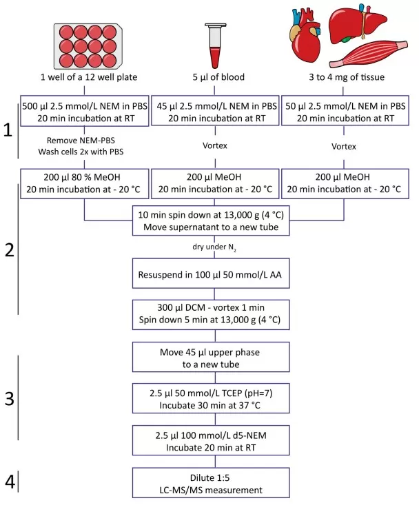
Figure 3. Two-step isotope-labeling workflow for determining the reduced-to-oxidized glutathione ratio by MRM-based LC-MS/MS. Reproduced from Tomin et al. (2020), Metabolites, 10(2), 71, under the Creative Commons Attribution 4.0 International License (CC BY 4.0).
Abbreviations: NEM, N-ethylmaleimide; d5-NEM, deuterated N-ethylmaleimide; TCEP, tris(2-carboxyethyl)phosphine; MRM, multiple reaction monitoring.
6. GSH/GSSG Analysis in Metabolomics and Multi-Omics
GSH/GSSG becomes more informative when interpreted within sulfur amino acid metabolism, NADPH supply, antioxidant enzymes, lipid oxidation, and phenotype. This framework helps distinguish substrate limitation, altered synthesis, increased oxidation, impaired regeneration, and downstream tissue injury.
6.1 Metabolic Context for GSH/GSSG Interpretation
Metabolic context can be built by profiling cysteine/cystine, glutamate, glycine, and γ-glutamylcysteine. These measurements can help distinguish precursor limitation from rapid oxidation or conjugation.
Depending on the research question, glutathione profiling may be combined with lipid-peroxidation products, GSH conjugates, or other pathway-specific metabolites.
6.2 Linking GSH/GSSG to Transcriptomic and Proteomic Regulation
Transcriptomic and proteomic data can connect metabolite changes to functional modules: GCLC/GCLM/GSS for synthesis, SLC7A11 for substrate acquisition, GSR/GPX/GST for regeneration and use, pentose phosphate pathway enzymes for NADPH supply, and NRF2-KEAP1 signaling for coordinated antioxidant adaptation.
In-parallel metabolomic and proteomic measurements can broaden the interpretation of glutathione-pathway activity (Wasinger et al., 2025). However, causal conclusions still require functional validation.
6.3 Selecting a GSH/GSSG Metabolomics Strategy
The analytical strategy should follow the research question. Targeted LC-MS/MS is suitable for focused hypotheses requiring rigorous stabilization and quantification, whereas discovery-oriented studies may combine broader profiling with targeted follow-up. Matrix, sample volume, stability, concentration range, and quantitative requirements should be evaluated before collection.
7. Frequently Asked Questions About GSH and GSSG
7.1 What Is the Main Difference Between GSH and GSSG?
GSH is the reduced form with a free cysteinyl thiol, whereas GSSG is the oxidized form created when two GSH molecules form a disulfide bond. Both are normal, interconvertible components of the same glutathione redox system.
7.2 What Does a Low GSH/GSSG Ratio Indicate?
A lower GSH/GSSG ratio generally indicates a more oxidized glutathione pool, but it does not identify the cause. Increased oxidation, impaired GSSG reduction, limited NADPH regeneration, reduced synthesis, and preanalytical artifacts can produce similar changes.
7.3 Why Can Sample Handling Distort GSH/GSSG Results?
GSH can oxidize after collection, while GSSG is present at much lower concentrations. Delayed processing, inadequate cooling, hemolysis, repeated freeze-thaw cycles, or insufficient thiol stabilization can lower measured GSH and falsely elevate GSSG. Rapid, standardized handling is therefore essential (Tomin et al., 2020; Thiel et al., 2023).
7.4 Is the GSH/GSSG Ratio the Same as Glutathione Redox Potential?
No. The ratio describes the relative distribution of GSH and GSSG, whereas redox potential also depends on their absolute concentrations, pH, and temperature. The ratio is useful for controlled comparisons but is not a complete thermodynamic description of redox state.
How MetwareBio Supports GSH/GSSG and Redox Metabolomics Research
Accurate GSH/GSSG analysis depends on matching sample collection, stabilization, analytical platform, and quantitative strategy to the biological question. MetwareBio supports LC-MS-based metabolite analysis, quality control, and pathway-level interpretation for glutathione-related and sulfur amino acid research.
Contact the MetwareBio team to discuss sample type, stabilization requirements, quantitative goals, and a study-specific analytical workflow.
Contact UsRead More: Redox Metabolism and Multi-Omics Strategies
These articles extend the GSH/GSSG discussion to related metabolic pathways, analytical methods, and multi-omics integration strategies for studying oxidative stress and redox biology in disease research.
Connects oxidative stress to lipid peroxidation products and human disease, extending the GSH/GSSG discussion to downstream lipid oxidation markers and their clinical relevance across inflammatory and metabolic conditions.
Explores how methionine metabolism supplies cysteine through the transsulfuration pathway, directly linking one-carbon metabolism to glutathione synthesis and cellular redox balance in the context of the GSH/GSSG system.
Details MetwareBio's enhanced targeted metabolomics platform for energy metabolism, including NADPH-related metabolites that directly feed glutathione regeneration and redox homeostasis.
Demonstrates how multi-omics integration strengthens disease research, complementing the GSH/GSSG framework with proteomic and lipidomic context for metabolic and inflammatory stress conditions.
Outlines practical strategies for combining proteomic and metabolomic data, directly relevant to linking GSH/GSSG measurements with antioxidant enzyme expression and pathway regulation in redox research.
Covers statistical methods for identifying differential metabolites, applicable to GSH/GSSG ratio analysis and biomarker validation across disease models and treatment conditions.
References
- Al-Temimi, A. A., Al-Mossawi, A.-E.-B., Al-Hilifi, S. A., Korma, S. A., Esatbeyoglu, T., Rocha, J. M., & Agarwal, V. (2023). Glutathione for food and health applications with emphasis on extraction, identification, and quantification methods: A review. Metabolites, 13(4), 465. https://doi.org/10.3390/metabo13040465
- Aoyama, K. (2021). Glutathione in the brain. International Journal of Molecular Sciences, 22(9), 5010. https://doi.org/10.3390/ijms22095010
- Fujii, J., Ito, J., Zhang, X., & Kurahashi, T. (2011). Unveiling the roles of the glutathione redox system in vivo by analyzing genetically modified mice. Journal of Clinical Biochemistry and Nutrition, 49(2), 70–78. https://doi.org/10.3164/jcbn.10-138SR
- Jiang, X., Stockwell, B. R., & Conrad, M. (2021). Ferroptosis: Mechanisms, biology and role in disease. Nature Reviews Molecular Cell Biology, 22, 266–282. https://doi.org/10.1038/s41580-020-00324-8
- Jones, D. P. (2002). Redox potential of the GSH/GSSG couple: Assay and biological significance. Methods in Enzymology, 348, 93–112. https://doi.org/10.1016/S0076-6879(02)48630-2
- Jyotsana, N., Ta, K. T., & DelGiorno, K. E. (2022). The role of cystine/glutamate antiporter SLC7A11/xCT in the pathophysiology of cancer. Frontiers in Oncology, 12, 858462. https://doi.org/10.3389/fonc.2022.858462
- Lu, S. C. (2013). Glutathione synthesis. Biochimica et Biophysica Acta - General Subjects, 1830(5), 3143–3153. https://doi.org/10.1016/j.bbagen.2012.09.008
- Thiel, A., Weishaupt, A.-K., Nicolai, M. M., Lossow, K., Kipp, A. P., Schwerdtle, T., & Bornhorst, J. (2023). Simultaneous quantitation of oxidized and reduced glutathione via LC-MS/MS to study the redox state and drug-mediated modulation in cells, worms and animal tissue. Journal of Chromatography B, 1225, 123742. https://doi.org/10.1016/j.jchromb.2023.123742
- Tomin, T., Schittmayer, M., & Birner-Gruenberger, R. (2020). Addressing glutathione redox status in clinical samples by two-step alkylation with N-ethylmaleimide isotopologues. Metabolites, 10(2), 71. https://doi.org/10.3390/metabo10020071
- Wasinger, V. C., Bustamante, S., Najib, N., Diwan, A., Jayasena, T., Chowdhury, N. S., Beretov, J., & Schabrun, S. (2025). Enzymes drive glutathione shunt to explain oxidative state using an in-parallel multi-omic method. International Journal of Molecular Sciences, 26(8), 3632. https://doi.org/10.3390/ijms26083632