Lipids are a chemically diverse group of hydrophobic or amphipathic molecules that include fatty acids, glycerolipids, glycerophospholipids, sphingolipids, and sterols. They form biological membranes, store metabolic energy, and act as signaling molecules or signaling precursors. These functions are essential for maintaining cellular homeostasis, and disturbances in lipid metabolism are closely linked to metabolic dysfunction, inflammation, and disease. This article discusses how different lipid classes contribute to membrane architecture, energy storage, and signal transduction, with ceramide-S1P and oxylipin signaling as representative examples. It also explains how lipidomics can reveal lipid remodeling and provide molecular insights into lipid function and disease-related changes.
1. Lipid Diversity and Major Classes: The Basis of Cellular Functions
Lipids comprise several chemically distinct groups, and this structural diversity underlies their wide range of cellular functions. The LIPID MAPS classification system organizes lipids into eight categories: Fatty Acyls, Glycerolipids, Glycerophospholipids, Sphingolipids, Sterol Lipids, Prenol Lipids, Saccharolipids, and Polyketides (Fahy et al., 2009). Table 1 summarizes the defining structural features, representative examples, and major biological roles of these categories.
Table 1. Eight LIPID MAPS Lipid Categories: Structural Features, Representative Examples, and Major Biological Roles
| LIPID MAPS category | Core structural feature | Representative examples | Major biological roles |
|---|---|---|---|
| Fatty Acyls [FA] | Hydrocarbon chain with a carboxyl-derived head group; includes oxidized and conjugated derivatives. | Fatty acids; eicosanoids/oxylipins | Fuel and lipid precursors; local signaling mediators. |
| Glycerolipids [GL] | Glycerol linked to one or more acyl/alkyl chains, generally without a phosphate headgroup. | MAG; DAG; TAG | Energy storage and lipid intermediates; DAG signaling. |
| Glycerophospholipids [GP] | Glycerol with hydrophobic chains and a phosphate-containing polar headgroup. | PC; PE; PS; PI | Membrane structure, curvature and charge; signaling-precursor pools. |
| Sphingolipids [SP] | Sphingoid-base backbone, commonly with an amide-linked fatty acid and variable headgroups. | Sphingomyelin; ceramide; S1P | Membrane organization, recognition, stress responses, and signaling. |
| Sterol Lipids [ST] | Fused four-ring sterol/steroid nucleus with variable side chains and oxidation states. | Cholesterol; bile acids; steroid hormones | Membrane order; endocrine, bile-acid, and oxysterol signaling. |
| Prenol Lipids [PR] | Constructed from repeating isoprene units. | Ubiquinone; dolichol; retinoids | Electron transport, protein glycosylation, cofactors, and signaling. |
| Saccharolipids [SL] | Fatty acyls attached directly to a sugar-based backbone. | Lipid A; acylaminosugars | Bacterial envelope structure; host-microbe and innate immune interactions. |
| Polyketides [PK] | Products assembled by iterative condensation of acyl-derived building blocks. | Erythromycin; aflatoxin B1 | Bioactive natural products with ecological, pharmacological, or toxic effects. |
For the themes developed below, Fatty Acyls, Glycerolipids, Glycerophospholipids, Sphingolipids, and Sterol Lipids are especially relevant because they dominate membrane structure, energy storage, and many lipid-signaling pathways in mammalian cells. Prenol Lipids, Saccharolipids, and Polyketides broaden the classification to other isoprenoid-, sugar-, and polyketide-derived lipids. The following sections focus on the classes most directly involved in membrane architecture, storage, and signaling.
2. Membrane Lipids and Cell Membrane Architecture
Biological membranes are amphipathic lipid bilayers whose hydrophobic core restricts uncontrolled passage of ions and most polar solutes while allowing lateral reorganization of lipids and proteins. Their composition sets bilayer thickness, packing, curvature, surface charge, and the local environment experienced by membrane proteins, shaping transport, remodeling, trafficking, and signaling-complex assembly.
The two plasma-membrane leaflets are also compositionally distinct. Experimental mapping has shown asymmetry in headgroups, acyl-chain unsaturation, and packing, giving proteins on the two membrane faces different physical environments (Lorent et al., 2020).
2.1 Membrane Fluidity, Curvature, and Lipid Packing
Acyl-chain length and unsaturation influence how tightly lipids pack: longer and more saturated chains generally increase order, whereas cis double bonds introduce kinks that loosen packing. Cholesterol can increase membrane bending rigidity and alter packing in a composition-dependent way rather than acting as a universal “fluidizer” (Doole et al., 2022). Headgroup size and molecular shape also affect spontaneous curvature, which becomes especially important when membranes bud, fuse, or form highly curved tubules.
These effects operate at the species level, not just the class level. Two samples with similar total phosphatidylcholine or phosphatidylethanolamine abundance can still have different membrane behavior if chain length or unsaturation shifts. This is one reason species-level lipidomics is more informative for membrane-remodeling studies than a single total-phospholipid measurement.
2.2 Membrane Lipid Organization, Protein Recruitment, and Trafficking
Membrane composition also controls which proteins can accumulate at a given site. Phosphatidylinositol itself is unevenly distributed across intracellular membranes (Zewe et al., 2020), while phosphoinositides provide compartment-specific cues that recruit proteins involved in membrane traffic and contact-site organization (Posor et al., 2022). Cholesterol, sphingolipids, and anionic phospholipids further influence local packing, electrostatics, and receptor organization. Rather than behaving as permanent “rafts,” many membrane nanodomains are transient and reorganize with trafficking, metabolism, and receptor activity (Bernardino de la Serna et al., 2016). Figure 1 summarizes several of the membrane properties that underlie this organization.
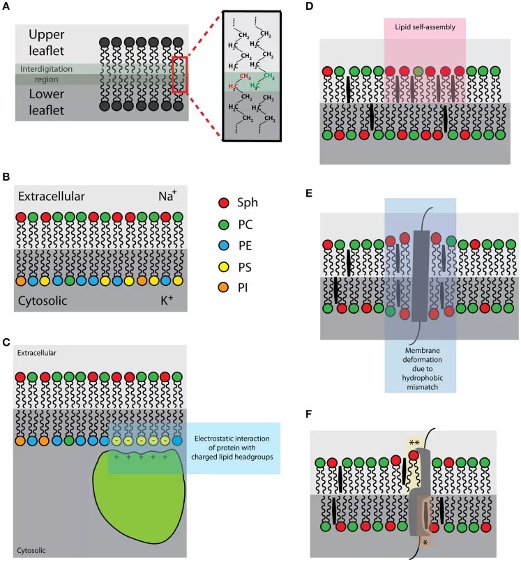
Figure 1. Intrinsic membrane properties that shape lipid organization and protein behavior, including leaflet asymmetry, lipid self-assembly, hydrophobic mismatch, and lipid-protein interactions. Image reproduced from Figure 2 in Bernardino de la Serna et al. (2016), Frontiers in Cell and Developmental Biology, under CC BY 4.0.
When membrane remodeling is expected to differ across tissue regions, MetwareBio Untargeted Spatial Lipidomics can complement bulk measurements by using MALDI mass spectrometry imaging to map lipid distributions directly in tissue sections.
3. Lipid Droplets, Energy Storage, and Metabolic Buffering
Cells package neutral lipids into lipid droplets instead of dispersing them through the aqueous cytosol. This packaging solves two problems at once: it concentrates energy-rich material and buffers cells against excess free fatty acids that could otherwise perturb membranes or metabolism.
3.1 Triacylglycerol Storage and Fatty-Acid Mobilization
TAGs are well suited to energy storage because their fatty-acyl chains are highly reduced and can be packed without the large hydration shell required by many soluble metabolites. Lipid droplets store TAGs together with cholesteryl esters in a neutral-lipid core surrounded by a phospholipid monolayer. When demand rises, lipolysis and lipophagy release fatty acids that can enter mitochondrial β-oxidation or be reused for membrane and signaling-lipid synthesis (Mathiowetz & Olzmann, 2024). Storage is reversible: the droplet is a regulated reservoir, not a terminal sink.
Direct cell-biological studies have shown fatty-acid transfer from lipid droplets toward mitochondria during nutrient limitation, linking mobilization to oxidation at the organelle level (Wang et al., 2021). This physical coordination helps explain why droplet abundance by itself says little about whether stored lipid is being actively used.
3.2 Lipid Droplets as Metabolic Buffers and Organelle Contact Sites
Lipid droplets buffer fluctuations in fatty-acid supply. Esterifying fatty acids into TAG reduces the pool of unesterified fatty acids available to perturb membranes or generate bioactive intermediates, while keeping those carbons available for later use. Droplets form functional contacts with the endoplasmic reticulum, mitochondria, and peroxisomes, supporting lipid exchange between storage, synthesis, and oxidation pathways (Mathiowetz & Olzmann, 2024).
A larger TAG pool may therefore reflect active buffering during lipid excess, reduced mobilization, increased synthesis, reduced oxidation, or several processes at once. Two samples can even have similar TAG abundance while differing substantially in synthesis and turnover. Quantitative lipidomics can define which TAG and cholesteryl-ester species change, but flux or turnover questions require time-resolved or isotope-based experiments beyond a steady-state lipid profile.
4. Lipid Signaling: Membrane Precursors and Bioactive Mediators
Lipid signaling is tightly coupled to membrane metabolism. Some signals are generated within seconds from membrane precursors, others emerge through enzymatic interconversion of lipid pools, and some are released to act on neighboring or distant cells. Interpretation hinges on where a lipid was produced and which enzymes or receptors can respond to it, not simply on its measured abundance.
4.1 Phosphoinositide, DAG, and Phosphatidic Acid Signaling
Phosphoinositides are low-abundance membrane lipids whose phosphorylation patterns help define membrane identity and recruit proteins with specific lipid-binding domains (Posor et al., 2022). A classic example is phosphatidylinositol 4,5-bisphosphate [PI(4,5)P2]. Phospholipase C cleaves PI(4,5)P2 to generate membrane-retained DAG and soluble inositol 1,4,5-trisphosphate (IP3), coupling a membrane-lipid reaction to protein kinase C activation and calcium signaling. DAG is also a metabolic branch point: diacylglycerol kinases can phosphorylate it to phosphatidic acid, which influences membrane remodeling and signaling (Kim & Wang, 2020). One membrane reaction can therefore create a membrane-associated messenger, a soluble second messenger, and a downstream lipid intermediate with distinct cellular effects.
4.2 Ceramide-Sphingosine-S1P Signaling Network
Ceramide, sphingosine, and S1P are metabolically connected but do not form a simple one-way pathway. Ceramide can arise through de novo synthesis or turnover of complex sphingolipids; ceramidases generate sphingosine, and sphingosine kinases produce S1P. These reactions are spatially organized, and regulated transport between compartments helps create distinct local sphingolipid pools (Körner & Fröhlich, 2022). Figure 2 shows a simplified view of these connections.
Ceramide participates in membrane organization, stress responses, apoptosis-related pathways, and metabolic regulation. Its effects vary with acyl-chain composition and subcellular location (Green et al., 2021). S1P can act inside cells and can also be exported to activate five G-protein-coupled S1P receptors; structural work on S1PR1 and S1PR5 shows how receptor context helps determine downstream signaling (Yuan et al., 2021).
The “ceramide-S1P rheostat” is useful shorthand, but it is not a fixed rule that ceramide always promotes cell death while S1P always promotes survival. Molecular species, compartment, receptor subtype, tissue environment, and cell state can all shift the biological outcome (Green et al., 2021).
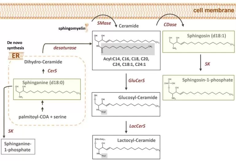
Figure 2. Ceramide-sphingosine-S1P metabolism within the sphingolipid pathway. The panel shows de novo ceramide synthesis, ceramide-to-sphingosine-to-S1P conversion, and related complex sphingolipids. Image cropped from Figure 1E in Neb et al. (2025), Frontiers in Immunology, licensed under CC BY 4.0; no panel content was altered.
4.3 PUFA-Derived Oxylipins and Intercellular Signaling
Polyunsaturated fatty acids (PUFAs) such as arachidonic acid, eicosapentaenoic acid, and docosahexaenoic acid can be released from membrane lipids and converted to oxygenated mediators. Oxylipins are the broader family; eicosanoids are a major subset that includes prostaglandins, leukotrienes, and thromboxanes. Many of these molecules are produced transiently and locally, so tissue or circulating abundance may not identify the site of signaling. Production is only half of the equation: mitochondrial fatty-acid oxidation machinery can contribute to oxylipin clearance during inflammatory activation (Misheva et al., 2022). A measured increase may reflect faster synthesis, slower removal, or both.
When oxylipins are the main biological question, a focused assay can be more informative than treating them as a small part of a broad lipid screen. MetwareBio Oxylipins Targeted Metabolomics provides LC-MS/MS-based absolute quantification for 141 predefined oxylipins, supporting pathway-focused inflammation, cardiovascular, and lipid-mediator research.
5. Lipid Dysregulation in Disease and Translational Research
Lipid dysregulation is informative in disease because membrane composition, storage capacity, and signaling often change together. The useful question is rarely whether lipids are simply “high” or “low,” but which species and pathways have shifted and whether those changes occur in the relevant tissue context. This makes lipidomics useful for biomarker discovery and mechanism studies when molecular patterns are linked to phenotype.
5.1 Cardiometabolic Disease and Cardiovascular Risk
Cardiometabolic disease illustrates why conventional lipid measurements and lipidomics answer different questions. Obesity, insulin resistance, fatty liver disease, and cardiovascular disease can involve TAG storage, fatty-acid handling, phospholipid remodeling, sphingolipid metabolism, and cholesterol-derived metabolites simultaneously. Ceramides are a major research focus because individual species and ceramide-to-phosphatidylcholine patterns have been associated with cardiovascular risk, while their effects depend on acyl-chain structure and tissue context (Green et al., 2021; Klingenberg et al., 2025). A circulating signature, however, should not be treated as direct evidence of what is occurring in a specific tissue.
When the pathway is not yet known, MetwareBio Quantitative Lipidomics can semi-quantitatively profile more than 4,000 lipid species across 51 classes using UPLC-MS/MS, MRM acquisition, and class-specific internal standards. When cholesterol conversion or the gut-liver axis is central to the hypothesis, Bile Acid Targeted Metabolomics provides absolute quantification of 65 bile acids. The two workflows separate broad remodeling from focused pathway measurement rather than forcing both questions into one assay.
5.2 Cancer Metabolism and Tumor Lipid Remodeling
Tumor cells remodel lipid metabolism to support membrane synthesis, energy flexibility, redox adaptation, and signaling. Fatty-acid saturation can influence membrane and oxidative-stress responses, lipid droplets can buffer potentially damaging lipid species, and sphingolipid or phospholipid pathways can affect growth and stress tolerance. Reviews of metastatic cancer biology emphasize that lipid availability and composition can shape tumor-cell behavior and treatment response rather than acting only as passive metabolic readouts (Vogel et al., 2024).
Sample context is particularly important in tumors. Bulk lipidomics can define group-level remodeling, whereas Untargeted Spatial Lipidomics can map regional lipid distributions in tissue sections by MALDI-MSI when tumor, stromal, necrotic, or treatment-responsive regions may differ. For mechanism-oriented projects, integrating lipid changes with protein or transcript data can help prioritize enzymes, transporters, and signaling pathways for follow-up.
Similar principles apply in neurodegeneration and inflammatory disease, where cholesterol transport, sphingolipid metabolism, and lipid-mediator signaling can vary by cell type and tissue region. APOE-linked neurobiology is a clear example of how altered lipid handling can become disease relevant (Yang et al., 2023). When localization and mediator concentration are both important, spatial lipidomics and targeted oxylipin analysis answer complementary questions rather than interchangeable ones.
6. Lipidomics Strategies for Functional and Disease Research
A useful lipidomics design starts with the biological claim the study needs to support. Broad remodeling, precise quantification of a known pathway, tissue localization, and cross-omics mechanism are different questions. Structural annotation also has limits: a sum composition such as PC 34:1 reports total carbon number and unsaturation but not the individual fatty-acyl chains or their positions, so identification level should be reported when species-level biology is interpreted (McDonald et al., 2022).
6.1 Broad-Coverage Profiling and Targeted Lipid Quantification
Broad-coverage quantitative lipidomics is useful for discovery questions such as which classes and species shift together and which pathways deserve follow-up. Targeted assays are better suited to predefined pathways in which concentration accuracy is the priority. MetwareBio’s broad quantitative lipidomics is semi-quantitative across thousands of predefined lipid species, whereas targeted oxylipin and bile-acid assays use authentic standards and calibration curves for absolute quantification. Targeted panels gain analytical focus but cannot reveal compounds outside the panel.
6.2 Spatial Lipidomics and Multi-Omics Integration
Bulk lipidomics averages signals across the extracted sample and cannot show whether a lipid is concentrated at a tumor edge, inflammatory focus, or other tissue region. Spatial lipidomics preserves this anatomical information. Multi-omics integration addresses a different gap: proteomics or transcriptomics can indicate which enzymes and regulatory programs accompany lipid remodeling and help narrow mechanistic hypotheses. Cross-omics correlation strengthens biological context but does not by itself prove causality.
Table 2. Matching Lipid Research Questions to MetwareBio Analytical Workflows
| Research question | Recommended MetwareBio workflow | What it adds | Key interpretation boundary |
|---|---|---|---|
| Global lipid remodeling, membrane composition, storage, or biomarker discovery | Quantitative Lipidomics | Broad, semi-quantitative profiling of 4,000+ lipid species across 51 classes | Bulk abundance does not establish localization or flux |
| Inflammation and bioactive lipid mediators | Oxylipins Targeted Metabolomics | Absolute quantification of 141 predefined oxylipins by LC-MS/MS | Focused panel; concentration does not equal receptor or pathway activation |
| Cholesterol conversion and gut-liver signaling | Bile Acid Targeted Metabolomics | Absolute quantification of 65 bile acids | Pathway-specific measurement rather than a global lipidome |
| Tissue heterogeneity and local lipid remodeling | Untargeted Spatial Lipidomics | MALDI-MSI mapping of lipid distributions at 5–100 µm with a 2,900+ lipid database | Spatial signal is not the same as absolute concentration or metabolic flux |
| Cross-layer mechanism and pathway prioritization | Multi-Omics Integration | Connect lipid/metabolite changes with proteins, transcripts, pathways, and phenotype | Association across omics layers still requires experimental validation |
MetwareBio Solutions for Lipid Function and Disease Research
MetwareBio’s lipid portfolio supports different levels of biological resolution. Quantitative Lipidomics provides broad species-level profiling for membrane remodeling, lipid storage, disease-associated signatures, and pharmacological studies. For focused pathways, Oxylipins Targeted Metabolomics and Bile Acid Targeted Metabolomics provide absolute quantification of predefined inflammatory mediators and bile acids.
For tissue studies, Untargeted Spatial Lipidomics maps lipid distributions in intact sections, while Multi-Omics integration can connect lipid changes with proteomic, transcriptomic, or metabolomic context. These options are most useful when they are selected around a defined evidence gap rather than added as parallel assays by default.
A practical workflow may begin with broad lipid remodeling, move to targeted quantification when a pathway is known, add spatial analysis when location matters, and use cross-omics data when regulatory context is needed. MetwareBio specialists can help align coverage, sample type, quantitative strategy, and downstream analysis with the intended biological conclusion.
Contact UsRead More: Lipid Biology and Lipidomics
These articles extend the topics covered here, from lipid classification and quantitative lipidomics platforms to specific lipid signaling pathways and data interpretation strategies.
Overview of MetwareBio quantitative lipidomics platform, profiling 4,000+ lipid species across 51 classes using UPLC-MS/MS with class-specific internal standards for broad coverage.
Comprehensive lipidomics service options including quantitative lipidomics, spatial lipidomics, and targeted oxylipin and bile acid assays for different research questions.
Deep dive into ceramide metabolism, covering de novo synthesis, sphingolipid turnover, and the role of ceramide species in metabolic disease and cardiovascular risk.
LC-MS/MS-based absolute quantification of 141 predefined oxylipins, supporting inflammation, cardiovascular, and lipid-mediator research with calibrated concentration data.
A beginner's guide to lipid classification, covering the eight LIPID MAPS categories, structural features, biological roles, and analytical approaches for lipid research.
A practical guide to lipidomics data interpretation, from raw data processing through statistical analysis, lipid identification, and biological pathway mapping.
References
- Fahy, E., Subramaniam, S., Murphy, R. C., Nishijima, M., Raetz, C. R. H., Shimizu, T., Spener, F., van Meer, G., Wakelam, M. J. O., & Dennis, E. A. (2009). Update of the LIPID MAPS comprehensive classification system for lipids. Journal of Lipid Research, 50(Suppl), S9–S14. https://doi.org/10.1194/jlr.R800095-JLR200
- Lorent, J. H., Levental, K. R., Ganesan, L., Rivera-Longsworth, G., Sezgin, E., Doktorova, M., Lyman, E., & Levental, I. (2020). Plasma membranes are asymmetric in lipid unsaturation, packing and protein shape. Nature Chemical Biology, 16, 644–652. https://doi.org/10.1038/s41589-020-0529-6
- Doole, F. T., Kumarage, T., Ashkar, R., & Brown, M. F. (2022). Cholesterol stiffening of lipid membranes. Journal of Membrane Biology, 255(4–5), 385–405. https://doi.org/10.1007/s00232-022-00263-9
- Zewe, J. P., Miller, A. M., Sangappa, S., Wills, R. C., Goulden, B. D., & Hammond, G. R. V. (2020). Probing the subcellular distribution of phosphatidylinositol reveals a surprising lack at the plasma membrane. Journal of Cell Biology, 219(3), e201906127. https://doi.org/10.1083/jcb.201906127
- Posor, Y., Jang, W., & Haucke, V. (2022). Phosphoinositides as membrane organizers. Nature Reviews Molecular Cell Biology, 23, 797–816. https://doi.org/10.1038/s41580-022-00490-x
- Bernardino de la Serna, J., Schütz, G. J., Eggeling, C., & Cebecauer, M. (2016). There is no simple model of the plasma membrane organization. Frontiers in Cell and Developmental Biology, 4, 106. https://doi.org/10.3389/fcell.2016.00106
- Wang, J., Fang, N., Xiong, J., Du, Y., Cao, Y., & Ji, W.-K. (2021). An ESCRT-dependent step in fatty acid transfer from lipid droplets to mitochondria through VPS13D–TSG101 interactions. Nature Communications, 12, 1252. https://doi.org/10.1038/s41467-021-21525-5
- Mathiowetz, A. J., & Olzmann, J. A. (2024). Lipid droplets and cellular lipid flux. Nature Cell Biology, 26, 331–345. https://doi.org/10.1038/s41556-024-01364-4
- Kim, S.-C., & Wang, X. (2020). Phosphatidic acid: An emerging versatile class of cellular mediators. Essays in Biochemistry, 64(3), 533–546. https://doi.org/10.1042/EBC20190089
- Körner, C., & Fröhlich, F. (2022). Compartmentation and functions of sphingolipids. Current Opinion in Cell Biology, 74, 104–111. https://doi.org/10.1016/j.ceb.2022.01.006
- Green, C. D., Maceyka, M., Cowart, L. A., & Spiegel, S. (2021). Sphingolipids in metabolic disease: The good, the bad, and the unknown. Cell Metabolism, 33(7), 1293–1306. https://doi.org/10.1016/j.cmet.2021.06.006
- Yuan, Y., Jia, G., Wu, C., Wang, W., Cheng, L., Li, Q., Li, Z., Luo, K., Yang, S., Yan, W., Su, Z., & Shao, Z. (2021). Structures of signaling complexes of lipid receptors S1PR1 and S1PR5 reveal mechanisms of activation and drug recognition. Cell Research, 31, 1263–1274. https://doi.org/10.1038/s41422-021-00566-x
- Misheva, M., Kotzamanis, K., Davies, L. C., Tyrrell, V. J., Rodrigues, P. R. S., Benavides, G. A., Hinz, C., Murphy, R. C., Kennedy, P., Taylor, P. R., Rosas, M., Jones, S. A., McLaren, J. E., Deshpande, S., Andrews, R., Schebb, N. H., Czubala, M. A., Gurney, M., Aldrovandi, M., … O’Donnell, V. B. (2022). Oxylipin metabolism is controlled by mitochondrial β-oxidation during bacterial inflammation. Nature Communications, 13, 139. https://doi.org/10.1038/s41467-021-27766-8
- Neb, H., Roth, V., Roos, J., Bauer, T., Urbschat, A., Heinicke, U., Angioni, C., Steinhilber, D., Piesche, M., Ferreirós, N., Gurke, R., Geisslinger, G., Utech, E., Zacharowski, K., Meybohm, P., Paulus, P., Schmitt, E., & Maier, T. J. (2025). Analysis of fatty acid-derived lipids in critically ill patients after cardiac surgery yields novel pathophysiologically relevant mediators with possible relevance for systemic inflammatory reactions. Frontiers in Immunology, 15, 1148806. https://doi.org/10.3389/fimmu.2024.1148806
- Klingenberg, R., Leiherer, A., Dobrev, D., Kaski, J. C., Levkau, B., März, W., Sossalla, S., von Eckardstein, A., & Drexel, H. (2025). Ceramides in cardiovascular disease: emerging role as independent risk predictors and novel therapeutic targets. Cardiovascular Research, 121(9), 1345–1358. https://doi.org/10.1093/cvr/cvaf093
- Vogel, F. C. E., Chaves-Filho, A. B., & Schulze, A. (2024). Lipids as mediators of cancer progression and metastasis. Nature Cancer, 5, 16–29. https://doi.org/10.1038/s43018-023-00702-z
- Yang, L. G., March, Z. M., Stephenson, R. A., & Narayan, P. S. (2023). Apolipoprotein E in lipid metabolism and neurodegenerative disease. Trends in Endocrinology & Metabolism, 34(7), 430–445. https://doi.org/10.1016/j.tem.2023.05.005
- McDonald, J. G., Ejsing, C. S., Kopczynski, D., Holčapek, M., Aoki, J., Arita, M., Arita, M., Baker, E. S., Bertrand-Michel, J., Bowden, J. A., Brügger, B., Ellis, S. R., Fedorova, M., Griffiths, W. J., Han, X., Hartler, J., Hoffmann, N., Koelmel, J. P., Köfeler, H. C., … Ekroos, K. (2022). Introducing the Lipidomics Minimal Reporting Checklist. Nature Metabolism, 4(9), 1086–1088. https://doi.org/10.1038/s42255-022-00628-3