Cultured-cell systems provide controlled models for investigating how genotype, nutrient availability, drug treatment, and environmental conditions reshape cellular metabolism. However, the cellular metabolome is highly responsive to cell density, medium composition, nutrient depletion, viability, washing, detachment, and delays in metabolic quenching. These variables can alter intracellular metabolite levels before LC-MS analysis, making cell culture conditions and sample collection integral parts of experimental design in cellular metabolomics. This article explains how cellular metabolomics can be used to study intracellular and extracellular metabolism in cultured mammalian cells. It covers experimental design, sample collection for adherent and suspension cells, the selection of untargeted, targeted, or lipidomics strategies for different research objectives, data normalization and quality control. By connecting analytical choices with the biological questions, this guide helps researchers design more reproducible cell culture metabolomics studies and interpret metabolic changes with greater confidence.
1. What Is Cellular Metabolomics?
Cellular metabolomics is the systematic measurement and analysis of metabolites at the cellular level to characterize metabolic states and responses to biological or experimental perturbations. In conventional cell culture studies, the analysis is usually performed on populations of cells using mass spectrometry-based platforms such as LC-MS, with intracellular metabolites measured from harvested cells and extracellular metabolites assessed from the culture medium when relevant to the research question.
Because metabolites are direct participants in energy production, biosynthesis, redox regulation, signaling, and nutrient utilization, their abundance patterns provide a functional view of how cells respond to changes in genotype, nutrient availability, drug exposure, or environmental conditions. Cellular metabolomics is therefore particularly useful when the objective is to identify metabolic consequences of a perturbation, discover pathways associated with a cellular phenotype, characterize drug-induced metabolic responses, or generate hypotheses about altered substrate use and metabolic adaptation (Zhao et al., 2024). The resulting data describe the metabolic state captured at the time of sampling, so interpretation depends strongly on well-controlled culture conditions and rapid, standardized sample collection.
The term cellular metabolomics describes an analytical approach and should be distinguished from cellular metabolism, which refers to the underlying biochemical reactions and metabolic networks operating within cells. Metabolomics measures the resulting metabolite pools and patterns; it does not by itself provide a complete measurement of every metabolic reaction or its rate. Conventional cellular metabolomics should also be distinguished from single-cell metabolomics. Most cultured-cell metabolomics studies analyze pooled populations and therefore report an average metabolic profile across many cells, whereas single-cell metabolomics measures individual cells to investigate metabolic heterogeneity within a population. These approaches address different biological questions and have different requirements for sample handling, analytical sensitivity, and data interpretation.
2. What Does Cell Culture Metabolomics Measure?
Cell culture metabolomics measures metabolites in two main compartments: the cells themselves and the surrounding culture medium. These intracellular and extracellular measurements capture different aspects of the same metabolic system.
2.1 Intracellular Metabolomics: Cellular Metabolite Profiling
Intracellular metabolomics measures metabolites contained within harvested cells and provides a snapshot of the cellular metabolite pool at the time of sampling. The detected compounds may include intermediates from central carbon metabolism, amino acid and nucleotide metabolism, redox pathways, and lipid metabolism. Changes across related metabolites can reveal coordinated shifts in cellular biochemical networks, making intracellular profiling the primary approach for characterizing metabolic changes occurring within a cell population.
2.2 Extracellular Metabolomics: Metabolite Uptake and Release
Extracellular metabolomics analyzes metabolites in conditioned culture medium to characterize the chemical exchange between cells and their environment. Depletion of medium components can reflect cellular uptake, while accumulation of extracellular metabolites can reflect release or secretion. This readout is especially informative for nutrients such as glucose, glutamine, and amino acids and for products such as lactate or other secreted metabolites. Because the culture medium already contains nutrients and other metabolites before cells are introduced, appropriate cell-free or baseline medium controls are important for distinguishing cell-associated changes from the original medium composition (Balcerczyk et al., 2020).
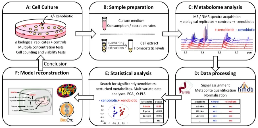
Figure 1. General workflow for metabolomics analysis of mammalian cell cultures, integrating extracellular medium analysis, intracellular metabolite extraction, analytical measurement, and downstream data analysis. Reproduced from Balcerczyk et al., 2020, International Journal of Molecular Sciences, 21, 6843. CC BY 4.0.
2.3 Integrating Intracellular and Extracellular Metabolomics
Paired intracellular and extracellular profiling can distinguish questions that are difficult to resolve from either compartment alone. An intracellular increase in a metabolite may be easier to interpret when the corresponding extracellular compound shows increased consumption or release. Likewise, a fall in intracellular abundance accompanied by greater extracellular accumulation can suggest enhanced export or leakage as a possibility that deserves follow-up. The combination therefore adds directionality to the biological reasoning, especially for nutrient exchange and secretory phenotypes. It still does not convert abundance measurements into reaction rates; rigorous flux conclusions require isotope-tracing designs or other kinetic measurements (Dong et al., 2022).
3. Experimental Design for Cellular Metabolomics
Cellular metabolomics is sensitive to biological and culture-related variation introduced before sample collection. Variables that are not part of the research question should therefore be standardized across experimental groups and documented throughout the study (Hayton et al., 2017).
3.1 Cell Model, Passage Number, Seeding Density, and Confluence
Cell source, passage history, seeding density, and confluence can influence growth state and the cellular metabolome. In practice, experiments should use the same cell source, a predefined passage range, and matched initial seeding densities across groups. Seeding density is best optimized in a pilot so that cells reach a comparable, non-overconfluent state at harvest. Cell number or confluence should also be recorded at collection, particularly when treatment affects proliferation. Seeding density has been shown to substantially alter metabolic profiles in cultured cells (Hayton et al., 2017; Ramirez-Hincapie et al., 2023).
3.2 Culture Medium and Environmental Conditions
Culture medium defines the nutrient environment in which cellular metabolism is measured. Medium formulation, glucose and glutamine concentrations, serum concentration and lot, culture volume, CO2, oxygen conditions, and medium-change schedule should remain consistent unless intentionally tested. Pilot experiments can confirm that major nutrients are not depleted before harvest, particularly in long incubations or high-density cultures. Medium composition itself can produce substantial metabolic differences, so culture conditions should be selected deliberately and reported clearly rather than treated as background information (Crooks et al., 2019; Vande Voorde et al., 2019).
3.3 Treatment Dose and Sampling Time
Treatment dose and sampling time determine the biological state captured by metabolomics. Dose and exposure duration should preferably be established in preliminary experiments, with viability and cell growth monitored in parallel. For studies focused on a specific metabolic response, sampling should generally occur before extensive nonspecific cell death develops, unless cytotoxicity is itself the intended endpoint. All replicates should follow the same interval from treatment to metabolic quenching. When the response is expected to change over time, predefined time-course sampling is more informative than combining samples collected at different stages (Bhinderwala and Powers, 2019).
3.4 Biological Replicates and Experimental Controls
Biological replicates should represent independently cultured samples exposed to the same experimental condition; repeated LC-MS injections of one extract do not substitute for biological replication. General metabolomics guidance commonly uses at least three biological replicates, with five or more preferred when feasible and variability is expected. Pilot data can support study-specific power planning. Controls should match the experimental design: untreated or vehicle controls are typically required for treatment studies, while cell-free medium controls are important when extracellular metabolites are analyzed. Cell viability and cell number should also be recorded when treatments substantially alter growth (Zhou et al., 2012).
4. Cell Harvesting and Metabolic Quenching for Cellular Metabolomics
Sample collection is a critical pre-analytical step in cellular metabolomics because intracellular metabolite levels can change rapidly after cells leave their controlled culture environment. A robust collection workflow therefore minimizes handling time, limits unnecessary washing, quenches metabolism as early as practical, and applies the same sequence and timing to every sample.
4.1 Principles of Metabolic Quenching
Metabolic quenching aims to stop ongoing biochemical reactions rapidly so that the measured metabolite profile reflects the intended sampling point. Temperature shifts, prolonged washing, delayed centrifugation, and repeated handling can alter labile intracellular metabolites or promote leakage. In practice, collection steps should be kept short, standardized across groups, and performed with preplanned reagents and equipment ready before harvest. Washing should be limited to what is necessary to remove extracellular medium without unnecessarily extending the collection process (Hayton et al., 2019).
4.2 Adherent Cell Collection for Metabolomics
A typical adherent-cell metabolomics workflow involves rapid medium removal, a brief wash when required, metabolic quenching, cell recovery, and immediate extraction or controlled freezing. Direct addition of cold extraction solvent can combine quenching and extraction, while other workflows quench first and recover cells by scraping. Enzymatic detachment with trypsin generally requires more handling before quenching and can alter metabolite recovery, so scraping or direct solvent-based collection is often preferred when compatible with the assay and normalization plan (Muschet et al., 2016; García-Cañaveras et al., 2016).
4.3 Suspension Cell Collection for Metabolomics
Suspension-cell metabolomics usually requires rapid separation of cells from the culture medium before intracellular extraction. Cells are typically collected by centrifugation, the supernatant is removed, and a brief wash may be included when extracellular contamination must be reduced. The resulting pellet is then quenched and extracted or rapidly frozen according to the validated workflow. Because metabolism continues during cell separation, centrifugation conditions and processing order should remain consistent across samples. For large experiments, staggered or balanced harvesting helps prevent systematic differences in collection time between experimental groups (Zhao et al., 2024).
4.4 Culture Supernatant Collection for Extracellular Metabolomics
Culture supernatant for extracellular metabolomics should be collected at a defined sampling time, ideally alongside the corresponding cellular sample. After collection, residual cells and debris are removed promptly and the clarified supernatant is processed or frozen under consistent conditions. Medium-only controls, and where appropriate fresh-medium or time-zero samples, provide the baseline needed to interpret changes in extracellular metabolites. When nutrient consumption or metabolite release is compared between groups, cell number or biomass should also be recorded so that differences in cell growth can be considered during data interpretation (Balcerczyk et al., 2020).
5. Choosing a Metabolomics Strategy for Cell Culture Studies
The appropriate metabolomics strategy depends primarily on the biological question and the metabolites of interest. Cultured-cell studies commonly use untargeted metabolomics for broad discovery, targeted metabolomics for predefined metabolites or pathways, and lipidomics when cellular lipid metabolism is the main focus.
5.1 Untargeted Metabolomics for Broad Metabolic Profiling
Untargeted metabolomics provides broad LC-MS profiling of metabolites and metabolic features without restricting analysis to a predefined panel. It is well suited to cultured-cell studies when the metabolic response is not fully known, allowing researchers to detect treatment-associated changes, identify candidate metabolites, and discover affected metabolic pathways. Metabolite annotation typically integrates accurate mass, retention time, MS/MS spectra, and reference standards where available, while relative abundance enables comparison across experimental groups (Alseekh et al., 2021).
5.2 Targeted Metabolomics for Pathway-Focused Analysis
Targeted metabolomics measures a predefined set of metabolites with optimized analytical conditions, providing focused and reproducible analysis of specific metabolic pathways. In cultured-cell studies, it is particularly suitable when the hypothesis centers on a defined metabolic pathway or metabolite class, such as central carbon metabolism, amino acid metabolism, organic acids, or other defined metabolite groups. Targeted LC-MS/MS methods can support relative, semi-quantitative, or absolute quantification depending on assay design and calibration. This strategy is commonly used for hypothesis-driven studies and for validating metabolic changes identified during discovery experiments (Turtoi et al., 2023).
5.3 Lipidomics for Cellular Lipid Remodeling
Lipidomics provides dedicated profiling of cellular lipids across classes such as phospholipids, sphingolipids, glycerolipids, and fatty-acid-related species. Specialized extraction, chromatographic separation, mass spectrometry, and lipid annotation improve coverage of the structural diversity of the cellular lipidome (García-Cañaveras et al., 2016). Lipidomics is therefore appropriate when a cultured-cell study focuses on membrane composition, lipid storage, lipid signaling, lipotoxicity, ferroptosis, or other forms of lipid remodeling. It can reveal coordinated changes across lipid classes that are difficult to characterize through general metabolomics alone.
6. Normalization and Quality Control in Cellular Metabolomics
Differences in cell number, biomass, extraction efficiency, and instrument response can affect measured metabolite signals independently of the biological condition. Cellular metabolomics therefore requires both appropriate sample normalization and analytical quality control to make metabolite profiles comparable across experimental groups. These steps should be incorporated into the study design and applied consistently throughout sample preparation, LC-MS analysis, and data processing.
6.1 Normalization Methods for Cell Metabolomics
Normalization adjusts metabolite measurements for differences in the amount of cellular material analyzed. Cell count is commonly used to express intracellular metabolite abundance per cell, while total protein or DNA can provide practical alternatives when direct counting is unavailable. The normalization factor should be measured consistently across all samples and selected according to the cell model and experimental design. When treatments substantially alter proliferation, cell size, or viability, these phenotypes should also be recorded to support interpretation of normalized metabolomics data (Muschet et al., 2016).
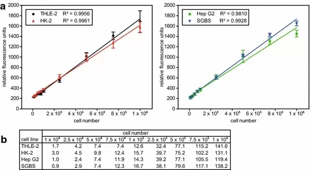
Figure 2. Relationship between DNA fluorescence and cell number across cultured mammalian cell lines, illustrating DNA-based normalization of cellular metabolomics samples. Reproduced from Muschet et al., 2016, Metabolomics, 12, 151. CC BY 4.0.
6.2 Analytical Quality Control in LC-MS Metabolomics
Analytical quality control evaluates the stability and reproducibility of the LC-MS metabolomics workflow. Pooled quality-control samples are commonly injected throughout the analytical sequence to monitor signal stability and instrument drift, while blank samples help identify background signals, contamination, and carryover. Internal standards can further track extraction and analytical performance, and randomized sample order reduces systematic bias between experimental groups and run position. For untargeted metabolomics, features with poor reproducibility or strong blank signals should be filtered before statistical and pathway analysis (Alseekh et al., 2021; Turtoi et al., 2023).
7. Interpreting Cellular Metabolomics: Metabolite Levels vs Metabolic Flux
Most cellular metabolomics studies measure steady-state metabolite abundance, providing a snapshot of intracellular metabolite pools at a defined sampling time. Changes in these pools can reveal altered metabolic states and help identify pathways associated with an experimental perturbation. However, metabolite concentration reflects the combined effects of synthesis, consumption, transport, and turnover, so changes in abundance do not directly measure the rate of metabolic reactions.
When the research question concerns substrate utilization, pathway routing, or metabolic flux, stable isotope tracing can provide additional information. Labeled substrates such as ^13C-glucose or ^13C-glutamine allow researchers to follow isotope incorporation into downstream metabolites and determine how specific nutrients contribute to metabolic pathways. Quantitative metabolic flux analysis may further incorporate labeling kinetics and mathematical modeling to estimate reaction rates (Dong et al., 2022).
8. Frequently Asked Questions About Cellular Metabolomics
What Types of Cell Samples Can Be Analyzed by Cellular Metabolomics?
Cellular metabolomics can be applied to established cell lines, cultured primary cells, stem-cell-derived or differentiated cells, and, with optimized low-input workflows, freshly isolated or FACS-purified cell populations. Cultured cells require careful control of medium composition, cell density, and harvesting conditions, while freshly isolated or sorted cells require rapid processing and sufficient analytical sensitivity to minimize ex vivo metabolic changes. This guide focuses on cultured mammalian cells, but the broader field also includes metabolomics of purified primary cell populations (Schönberger et al., 2023).
How Many Cells Are Needed for Cellular Metabolomics?
There is no universal cell-number requirement for cellular metabolomics because the required input depends on the cell type, cell size, metabolite abundance, analytical platform, and selected metabolomics strategy. Untargeted metabolomics, targeted metabolomics, and lipidomics may have different sample-input requirements, and low-abundance metabolites can require more starting material than abundant metabolic intermediates. Biological replicates must also be considered when planning the total number of cells needed for a study. Researchers should therefore confirm the sample requirements for the selected assay before the final cell culture experiment or sample collection begins.
How Should Cellular Metabolomics Be Designed When a Treatment Affects Cell Growth or Viability?
When a treatment alters cell proliferation or viability, cellular metabolomics should be designed to separate treatment-associated metabolic changes from differences caused by cell number or extensive cell death. Cell number, confluence, and viability should be recorded at harvest, and treatment dose and sampling time should be selected according to the biological objective. Appropriate normalization using cell count, protein, DNA, or another validated measure can improve comparability among samples. If strong growth inhibition or cytotoxicity is expected, multiple doses or time points can help distinguish early metabolic responses from later changes associated with loss of viability.
Planning a Cellular Metabolomics Study with MetwareBio
A successful cellular metabolomics study depends on coordinated decisions across experimental design, cell culture and sample collection, metabolite extraction, LC-MS analysis, and bioinformatics. MetwareBio supports this workflow by providing guidance on study design and cell sample collection, followed by end-to-end experimental and data-analysis services from metabolite extraction onward. Available options include untargeted metabolomics, a broad range of targeted metabolomics panels such as Central Carbon Metabolism and Amino Acid Metabolism, and lipidomics, enabling researchers to match the analytical approach to their specific metabolic question. If you are planning a cellular metabolomics project, contact MetwareBio to discuss your study design, sample strategy, and the most suitable analytical workflow.
Discuss Your Cell StudyRead More: Cellular Metabolomics, Sample Preparation, and LC-MS Workflow
These articles complement the current cellular metabolomics guide by detailing metabolomics strategy selection, cell sample preparation and normalization, LC-MS workflow quality control, and the targeted-pathway and lipidomics options available for cultured-cell studies.
Section 5 of this guide compares untargeted, targeted, and lipidomics strategies for cultured cells; this article expands that decision framework with a side-by-side look at analytical logic, quantification endpoints, sample constraints, cohort size, and staged discovery-then-verification designs.
Follows directly from the harvesting and quenching discussion in Section 4. It answers practical collection questions for cell samples, including how to prepare primary cells, recommended cell numbers, wash and snap-freeze steps, biological replicate recommendations, and normalization options when accurate cell counting is difficult.
Extends the analytical quality-control discussion in Section 6.2 with a five-step LC-MS workflow and its QA checkpoints, covering platform choice between triple quadrupole and high-resolution MS, internal-standard and blank controls, pooled QC injection frequency, retention-time drift monitoring, and matrix-effect mitigation.
Explains centering, scaling, and transformation methods, including auto scaling, Pareto scaling, and log transformation. These statistical preprocessing steps complement sample normalization by cell count, protein, or DNA, which addresses differences in cellular material.
An example of the pathway-focused strategy described in Section 5.2, providing absolute quantification of 80 metabolites across glycolysis, the TCA cycle, and the pentose phosphate pathway by LC-MS/MS in MRM mode, with project experience spanning cultured cells, tissues, and biofluids.
Background reading for the lipidomics option in Section 5.3. It covers lipid classification systems, the membrane, energy-storage, and signaling roles of lipids, how lipidomics differs from general metabolomics, and the analytical methods used for class-specific lipid profiling.
References
- Alseekh, S., Aharoni, A., Brotman, Y., et al. (2021). Mass spectrometry-based metabolomics: a guide for annotation, quantification and best reporting practices. Nature Methods, 18, 747-756. https://doi.org/10.1038/s41592-021-01197-1
- Balcerczyk, A., Damblon, C., Elena-Herrmann, B., et al. (2020). Metabolomic approaches to study chemical exposure-related metabolism alterations in mammalian cell cultures. International Journal of Molecular Sciences, 21(18), 6843. https://doi.org/10.3390/ijms21186843
- Bhinderwala, F., & Powers, R. (2019). NMR metabolomics protocols for drug discovery. Methods in Molecular Biology, 2037, 265-311. https://doi.org/10.1007/978-1-4939-9690-2_16
- Crooks, D. R., Fan, T. W.-M., & Linehan, W. M. (2019). Metabolic labeling of cultured mammalian cells for stable isotope-resolved metabolomics: Practical aspects of tissue culture and sample extraction. Methods in Molecular Biology, 1928, 1-27. https://doi.org/10.1007/978-1-4939-9027-6_1
- Dong, W., Rawat, E. S., Stephanopoulos, G., & Abu-Remaileh, M. (2022). Isotope tracing in health and disease. Current Opinion in Biotechnology, 76, 102739. https://doi.org/10.1016/j.copbio.2022.102739
- García-Cañaveras, J. C., López, S., Castell, J. V., Donato, M. T., & Lahoz, A. (2016). Extending metabolome coverage for untargeted metabolite profiling of adherent cultured hepatic cells. Analytical and Bioanalytical Chemistry, 408, 1217-1230. https://doi.org/10.1007/s00216-015-9227-8
- Hayton, S., Maker, G. L., Mullaney, I., & Trengove, R. D. (2017). Experimental design and reporting standards for metabolomics studies of mammalian cell lines. Cellular and Molecular Life Sciences, 74, 4421-4441. https://doi.org/10.1007/s00018-017-2582-1
- Hayton, S., Trengove, R. D., & Maker, G. L. (2019). Sample Preparation and Reporting Standards for Metabolomics of Adherent Mammalian Cells. In: High-Throughput Metabolomics: Methods and Protocols, Methods in Molecular Biology, 1978, 3-12. https://doi.org/10.1007/978-1-4939-9236-2_1
- Muschet, C., Möller, G., Prehn, C., et al. (2016). Removing the bottlenecks of cell culture metabolomics: fast normalization procedure, correlation of metabolites to cell number, and impact of the cell harvesting method. Metabolomics, 12, 151. https://doi.org/10.1007/s11306-016-1104-8
- Ramirez-Hincapie, S., Birk, B., Ternes, P., et al. (2023). A high-throughput metabolomics in vitro platform for the characterization of hepatotoxicity. Cell Biology and Toxicology, 39(6), 2899-2917. https://doi.org/10.1007/s10565-023-09809-6
- Schönberger, K., Mitterer, M., Glaser, K., et al. (2023). LC-MS-based targeted metabolomics for FACS-purified rare cells. Analytical Chemistry, 95(9), 4325–4334. doi: 10.1021/acs.analchem.2c04396.
- Turtoi, E., Jeudy, J., Henry, S., et al. (2023). Analysis of polar primary metabolites in biological samples using targeted metabolomics and LC-MS. STAR Protocols, 4(3), 102400. https://doi.org/10.1016/j.xpro.2023.102400
- Vande Voorde, J., Ackermann, T., Pfetzer, N., et al. (2019). Improving the metabolic fidelity of cancer models with a physiological cell culture medium. Science Advances, 5(1), eaau7314. https://doi.org/10.1126/sciadv.aau7314
- Zhao, Y., Sepehr, E., Vaught, C., Yourick, J., & Sprando, R. L. (2024). Cellular metabolomics: From sample preparation to high-throughput data analysis. Journal of Agriculture and Food Research, 15, 100935. https://doi.org/10.1016/j.jafr.2023.100935
- Zhou, B., Xiao, J. F., Tuli, L., & Ressom, H. W. (2012). LC-MS-based metabolomics. Molecular BioSystems, 8(2), 470-481. https://doi.org/10.1039/C1MB05350G