Short-chain fatty acids (SCFAs) connect gut microbial fermentation with intestinal and systemic metabolism, but their biological meaning depends on the sample being analyzed. Fecal concentrations describe the residual pool excreted after intestinal production, absorption, and transit, while plasma and serum capture circulating concentrations shaped by metabolite supply, host metabolism, and clearance. Selecting a matrix without considering these differences can lead researchers to confuse stool abundance with microbial production or miss a response in the relevant biological compartment. This article compares fecal, plasma, and serum SCFA analysis, explains the physiological and analytical factors that distinguish these matrices, and examines when paired sampling adds value. It provides a practical framework for choosing samples that match the research question and interpreting SCFA measurements within the biological context of each matrix.
1. Why SCFA Profiles Differ Across Sample Matrices
Intestinal short-chain fatty acids (SCFAs) are produced mainly through microbial fermentation of carbohydrates that escape digestion in the small intestine, with additional contributions from protein and amino acid fermentation. Acetate, propionate, and butyrate usually predominate, but their production patterns depend on substrate availability, microbial metabolic capacity, and cross-feeding between microorganisms. As intestinal contents move through the colon, changes in substrate supply and fermentation conditions create spatial and temporal variation in the luminal SCFA pool.
Most SCFAs are absorbed across the intestinal epithelium, where colonocytes utilize a substantial proportion of butyrate as an energy source. The remaining absorbed SCFAs enter the portal circulation and undergo further hepatic metabolism. Compound-specific differences in intestinal utilization and hepatic extraction result in greater systemic availability of acetate than of propionate or butyrate (Boets et al., 2017). Host metabolism also contributes to circulating acetate, while peripheral tissue uptake and clearance further shape blood concentrations. Meanwhile, absorption, microbial conversion, and intestinal transit continuously modify the SCFAs remaining in the lumen (Sakata, 2019).
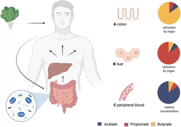
Figure 1. Production, distribution and yield of short-chain fatty acids (SCFAs) in different tissues. Image reproduced from Palm et al., 2022, Frontiers in Cardiovascular Medicine, licensed under CC BY 4.0. Original figure created with BioRender.com.
Different sample matrices therefore capture SCFAs at distinct points along the pathway from microbial production to absorption, host metabolism, and excretion. Sample selection determines which biological compartment is measured and, consequently, what an SCFA concentration can reveal about the underlying physiology.
2. Fecal SCFA Analysis
2.1 What Fecal SCFA Levels Represent
Fecal SCFA concentrations characterize the residual pool present in stool at collection. They reflect the net outcome of microbial production, absorption, conversion, and intestinal transit before excretion. This makes fecal analysis informative about the chemical environment represented in stool, although the measurement does not resolve how much SCFA was produced or absorbed separately (Sakata, 2019).
2.2 When to Choose Fecal SCFA Analysis
Fecal SCFAs are particularly useful when the study focuses on the intestinal metabolic environment. Examples include dietary-fiber or prebiotic interventions, microbiome studies that relate microbial composition or functional potential to fermentation products, and experiments asking whether an intervention shifts the relative profile of acetate, propionate, butyrate, or branched-chain fatty acids within stool. In these settings, the fecal matrix is biologically close to the microbial ecosystem of interest and can be collected noninvasively, which facilitates repeated sampling in longitudinal studies.
2.3 Limitations of Fecal SCFA Interpretation
Fecal SCFA concentrations are influenced by stool water content and intestinal transit as well as microbial metabolism. A change in concentration per gram of wet stool may partly reflect dilution or concentration of the sample, while altered transit changes the time available for fermentation and absorption. These variables should therefore be considered when relating fecal SCFA differences to dietary interventions or microbiome changes (Sakata, 2019).
3. Plasma and Serum SCFA Analysis
3.1 What Circulating SCFA Levels Represent
Plasma and serum SCFA concentrations reflect the balance between entry into circulation and removal by host tissues. Intestinal utilization and hepatic extraction limit the fraction of gut-derived SCFAs reaching peripheral blood, with compound-specific differences contributing to the predominance of acetate in circulation (Boets et al., 2017). Paired fecal and plasma measurements also show distinct SCFA profiles across the two matrices (Manokasemsan et al., 2024). Blood measurements are therefore useful for characterizing systemic exposure under the sampled conditions, although concentrations alone cannot resolve microbial production, absorption, or clearance rates.
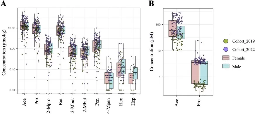
Figure 2. The individual SCFAs of healthy individuals (cohort 2019 + cohort 2022; N = 157) in fecal (A) samples and (B) plasma samples labeled by cohort. Image reproduced from Manokasemsan et al., 2024, Computational and Structural Biotechnology Journal, licensed under CC BY 4.0.
3.2 When to Choose Plasma or Serum SCFA Analysis
Blood SCFA analysis is well suited to studies in which the central question concerns host exposure or systemic physiology. Examples include nutritional or microbiome interventions designed to alter circulating microbial metabolites, studies relating SCFAs to metabolic or immune phenotypes measured in blood, and investigations of gut-host communication in which circulating SCFA concentrations are a prespecified endpoint. In these settings, plasma or serum places the measurement in the same physiological compartment as many clinical chemistry, cytokine, hormone, and metabolomics endpoints, which can simplify integrated analyses.
3.3 Plasma vs Serum for SCFA Measurement
Plasma and serum are prepared from blood using different collection and processing procedures. Plasma is collected with an anticoagulant and separated without clotting, whereas serum is obtained after coagulation. That difference can alter the chemical environment of the sample and introduces matrix-specific effects from anticoagulants, clotting, collection tubes, and processing time. For SCFA studies, the important design principle is consistency: plasma and serum should not be mixed within a study as though they were analytically interchangeable. Method-validation work has shown that tube chemistry can affect circulating SCFA analysis and has identified specific plasma and serum collection formats that perform more reliably under the evaluated GC-MS workflow (Bain et al., 2022). The exact preferred tube should therefore follow the validated analytical protocol used by the laboratory.
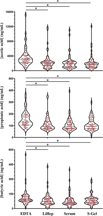
Figure 3. Violin plots to compare acetic acid (top), propionic acid (middle) and butyric acid (bottom) measurements from 51 healthy participants across different blood collection methods. Image reproduced from Bain et al., 2022, Journal of Mass Spectrometry and Advances in the Clinical Lab, licensed under CC BY 4.0.
4. Paired Fecal and Blood SCFA Analysis
Paired fecal and blood SCFA analysis helps researchers connect intestinal metabolic changes with circulating availability and host physiology. Integrating fecal SCFAs with microbiome data can identify microbial shifts associated with the stool metabolite response, while plasma or serum measurements establish whether a corresponding response is detectable in circulation. Relating both matrices to the same inflammatory, hormonal, or metabolic phenotypes then allows investigators to examine which compartment is more closely associated with the host response. Together, these measurements provide a connected view of microbial composition, intestinal metabolites, circulating SCFAs, and host outcomes.
Interpretation begins with the change in each SCFA within each matrix. In an intervention study, changes detected in both feces and blood indicate that the metabolic response extends across the two measured compartments. A fecal change without a detectable blood response indicates that the stool signal was not accompanied by a circulating signal under the study’s sampling conditions. Examining these patterns alongside host outcomes can help distinguish an association with the intestinal metabolic environment from one that tracks circulating SCFA availability. Cross-matrix correlations provide additional information about how closely the two measurements vary between individuals or across study visits.
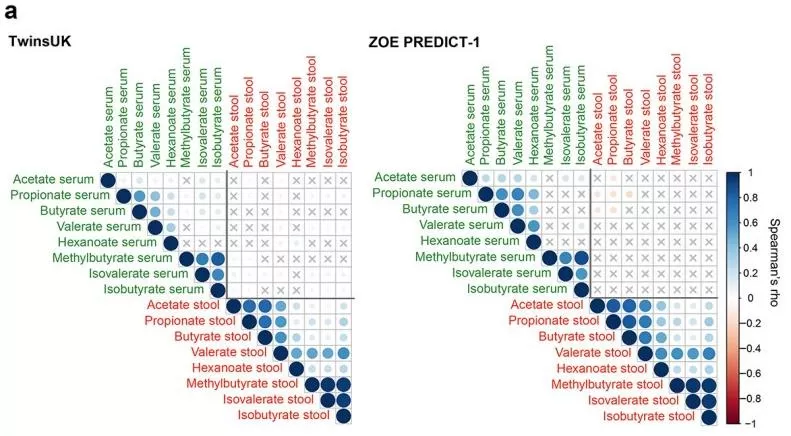
Figure 4. Fasting circulating and faecal SCFAs correlation in TwinsUK (n=2229) and ZOE PREDICT-1 (n=328). Image adapted from Nogal et al., 2023, Gut Microbes, licensed under CC BY 4.0.
Close agreement between fecal and blood profiles is not a prerequisite for useful integration. Farhat et al. (2021) reported low concordance for most fecal and serum SCFAs despite generally good technical reproducibility. This finding is consistent with absorption and host metabolism reshaping the relationship between intestinal and circulating pools, making divergent responses biologically informative when sampling and analytical quality are adequately controlled. Associations across these measurements can support a gut–host hypothesis, although establishing the causal pathway requires additional evidence.
5. Choosing Between Fecal and Blood SCFA Analysis
SCFA sample selection should follow the primary biological endpoint. Fecal analysis is appropriate for characterizing the residual intestinal SCFA pool in studies of diet and the gut microbiome. Plasma or serum analysis addresses circulating availability and its relationship to systemic host phenotypes. When a study aims to evaluate both intestinal and circulating responses, paired sampling provides the measurements needed to examine each compartment and their relationship.
| Selection criterion | Fecal SCFA analysis | Blood SCFA analysis | Paired fecal and blood SCFA analysis |
|---|---|---|---|
| Biological pool measured | Residual SCFAs in stool after intestinal production, absorption, metabolism, and transit | Circulating SCFAs shaped by absorption, host metabolism, tissue uptake, and clearance | Both residual fecal and circulating SCFA pools in the same participants |
| Primary research endpoint | Differences or changes in fecal SCFA concentrations and composition | Differences or changes in circulating SCFA concentrations and associations with host phenotypes | Responses in both compartments and the relationship between intestinal and circulating profiles |
| Typical applications | Dietary fiber, prebiotic, and microbiome studies focused on intestinal metabolic responses | Nutritional or microbiome studies focused on circulating metabolites, metabolic regulation, or immune phenotypes | Intervention studies examining whether intestinal metabolic changes are accompanied by circulating changes and how each relates to host outcomes |
| Complementary measurements | Microbiome composition and functional potential, dietary intake, stool consistency, and intestinal transit | Clinical chemistry, inflammatory markers, hormones, and systemic metabolomics | Integrated microbiome, dietary, systemic metabolomics, and host phenotype data |
| Main interpretive limitation | Concentrations reflect the balance between production and disappearance from the intestinal lumen; they do not directly quantify microbial production | Concentrations reflect the balance between systemic appearance and removal; they do not directly quantify colonic production or absorption | Paired concentrations alone cannot quantify transfer between compartments or establish a causal gut–host pathway |
6. Key Considerations for SCFA Analysis
6.1 Sample Collection and Storage
Preanalytical handling can affect SCFA measurements through sample heterogeneity, ongoing microbial activity, and analyte loss. Fecal collection should use consistent subsampling, homogenization, and storage procedures that minimize these effects. For plasma and serum, collection tube chemistry, clotting conditions, and the interval before separation should follow the laboratory’s validated protocol (Bain et al., 2022). Apply the chosen procedure consistently across groups and study visits, and record deviations that could affect interpretation. For archived specimens, assess whether processing or storage differences coincide with the biological groups being compared.
6.2 Matrix Effects and Analytical Sensitivity
Feces, plasma, and serum differ substantially in matrix composition and expected SCFA concentration, so one analytical workflow cannot be assumed to perform identically in all three. Modern GC-MS methods have been validated across multiple SCFA matrices, but those studies explicitly evaluate extraction recovery, matrix effects, precision, and calibration behavior rather than relying on a single generic preparation (Kim et al., 2022). Plasma and serum are particularly demanding because lower endogenous concentrations increase the importance of background contamination, extraction efficiency, and instrument sensitivity (Yao et al., 2022).
Absolute quantification requires a calibration strategy that accounts for SCFAs already present in biological samples. A surrogate matrix can be used when its analytical response, extraction recovery, and matrix effects have been evaluated against the target biological matrix. Isotope-labeled internal standards serve a complementary role in correcting extraction and analytical variability; they do not, by themselves, eliminate endogenous analyte background or establish the suitability of the calibration matrix (Kim et al., 2022).
6.3 Cross-Matrix Quantification and Interpretation
Absolute concentrations from feces and blood should not be compared as if they were measurements on a common scale. Fecal data are typically expressed relative to sample mass, whereas plasma and serum are reported per unit volume, and the biological compartments have different water content, transport dynamics, and metabolic turnover. A numerically larger fecal concentration therefore does not imply greater biological exposure than a lower plasma concentration. Interpretation should focus on within-matrix differences, compound-specific patterns, and the biological relationship between compartments.
7. SCFA Sample Selection FAQs
Do fecal SCFA levels reflect microbial production?
Fecal SCFA concentrations reflect the residual stool pool, shaped by microbial production, absorption, transit, and stool water content. They do not directly measure production rate. Estimating in vivo production requires additional kinetic information, such as stable-isotope measurements interpreted using an appropriate model (Boets et al., 2015; Sakata, 2019).
Can plasma and serum be used interchangeably for SCFA analysis?
They should not be treated as automatically interchangeable. Plasma is collected in the presence of an anticoagulant, whereas serum is produced after clotting, so the two matrices differ in preanalytical processing and can respond differently to tube chemistry and sample handling. Either matrix can be used when it is supported by the analytical method, but a study should keep the matrix, tube type, processing interval, and storage protocol consistent across all samples (Bain et al., 2022).
Should paired fecal and blood samples be collected at the same time?
When the objective is to relate the intestinal and systemic SCFA compartments, temporally matched collection is preferable because diet, treatment, intestinal transit, and other exposures can change over time. Exact simultaneous collection is not always practical, especially for stool, but the sampling window should be defined in advance and applied consistently. The analysis plan should also recognize that fecal material integrates intestinal events over a different time scale than a blood draw.
Can fecal and blood SCFA concentrations be compared directly?
Direct numerical comparison is generally not meaningful. Fecal and blood SCFAs are measured in different biological compartments, commonly use different concentration units, and have very different abundance ranges. The informative comparison is whether each matrix changes across groups or time points and whether paired values are associated after accounting for their distinct biological meaning. Low fecal-blood concordance can be expected even when both measurements are analytically reproducible (Farhat et al., 2021).
SCFA Analysis Services at MetwareBio
At MetwareBio, our SCFA analysis service provides absolute quantification of 17 short-chain, branched-chain, and selected medium-chain fatty acids using GC-MS/MS. Gas chromatographic separation and triple-quadrupole MRM enable selective measurement of structurally related fatty acids, while analyte-specific calibration, internal standards, and rigorous quality control support reliable concentration measurements. We analyze feces, intestinal contents, plasma, and serum using matrix-appropriate workflows for intestinal, circulating, or paired SCFA studies. If you need SCFA analysis for your research, please contact us to discuss your objectives and sample requirements.
Discuss Your SCFA ProjectRead More: SCFA Analysis and Sample Matrix Selection
These articles extend the topics above—from choosing between serum, plasma, feces, and other matrices for metabolomics, to the biology and measurement of individual SCFAs in gut–host research.
Plasma and serum differ in coagulation, anticoagulant chemistry, and sample processing—variables that directly shape measured metabolite profiles. This guide complements the plasma-vs-serum considerations above with metabolomics-specific study design advice.
A practical framework for matching sample matrix to research question, covering cells, tissues, blood components, urine, and other biofluids—the same matrix-choice logic applied to SCFA studies in this article.
Reviews how SCFAs are produced, how they act on host physiology, and how targeted SCFA panels support research—useful background for interpreting acetate, propionate, and butyrate data from any matrix.
Covers branched SCFA biology and the analytical pitfalls of measuring low-abundance fatty acids, including GC-MS vs LC-MS/MS choices and preanalytical confounders that echo the key considerations section above.
Explains how gut microbes produce SCFAs and how they influence immunity, metabolism, and disease—context for why fecal and circulating pools can tell different biological stories.
Explore BHB as a host-derived ketone body, including its metabolism, signaling roles, and measurement. A complementary perspective on how biological context and sample selection shape metabolite interpretation.
References
- Bain, J. T., Taal, M. W., Selby, N. M., Reynolds, J. C., & Heaney, L. M. (2022). Simple, high-throughput measurement of gut-derived short-chain fatty acids in clinically relevant biofluids using gas chromatography-mass spectrometry. Journal of Mass Spectrometry and Advances in the Clinical Lab, 25, 36-43. https://doi.org/10.1016/j.jmsacl.2022.07.002
- Boets, E., Deroover, L., Houben, E., Vermeulen, K., Gomand, S. V., Delcour, J. A., & Verbeke, K. (2015). Quantification of in vivo colonic short chain fatty acid production from inulin. Nutrients, 7(11), 8916-8929. https://doi.org/10.3390/nu7115440
- Boets, E., Gomand, S. V., Deroover, L., Preston, T., Vermeulen, K., De Preter, V., Hamer, H. M., Van den Mooter, G., De Vuyst, L., Courtin, C. M., Annaert, P., Delcour, J. A., & Verbeke, K. (2017). Systemic availability and metabolism of colonic-derived short-chain fatty acids in healthy subjects: a stable isotope study. The Journal of Physiology, 595(2), 541-555. https://doi.org/10.1113/JP272613
- Farhat, Z., Sampson, J. N., Hildesheim, A., Safaeian, M., Porras, C., Cortes, B., Herrero, R., Romero, B., Vogtmann, E., Sinha, R., & Loftfield, E. (2021). Reproducibility, temporal variability, and concordance of serum and fecal bile acids and short chain fatty acids in a population-based study. Cancer Epidemiology, Biomarkers & Prevention, 30(10), 1875-1883. https://doi.org/10.1158/1055-9965.EPI-21-0361
- Kim, K.-S., Lee, Y., Chae, W., & Cho, J.-Y. (2022). An improved method to quantify short-chain fatty acids in biological samples using gas chromatography-mass spectrometry. Metabolites, 12(6), 525. https://doi.org/10.3390/metabo12060525
- Manokasemsan, W., Jariyasopit, N., Poungsombat, P., Kaewnarin, K., Wanichthanarak, K., Kurilung, A., Duangkumpha, K., Limjiasahapong, S., Pomyen, Y., Chaiteerakij, R., Tansawat, R., Srisawat, C., Sirivatanauksorn, Y., Sirivatanauksorn, V., & Khoomrung, S. (2024). Quantifying fecal and plasma short-chain fatty acids in healthy Thai individuals. Computational and Structural Biotechnology Journal, 23, 2163-2172. https://doi.org/10.1016/j.csbj.2024.05.007
- Nogal, A., Asnicar, F., Vijay, A., Kouraki, A., Visconti, A., Louca, P., Wong, K., Baleanu, A.-F., Giordano, F., Wolf, J., Hadjigeorgiou, G., Davies, R., Michelotti, G. A., Franks, P. W., Berry, S. E., Falchi, M., Ikram, A., Ollivere, B. J., Zheng, A., … Menni, C. (2023). Genetic and gut microbiome determinants of SCFA circulating and fecal levels, postprandial responses and links to chronic and acute inflammation. Gut Microbes, 15(1), 2240050. https://doi.org/10.1080/19490976.2023.2240050
- Palm, C. L., Nijholt, K. T., Bakker, B. M., & Westenbrink, B. D. (2022). Short-chain fatty acids in the metabolism of heart failure—Rethinking the fat stigma. Frontiers in Cardiovascular Medicine, 9, 915102. https://doi.org/10.3389/fcvm.2022.915102
- Sakata, T. (2019). Pitfalls in short-chain fatty acid research: A methodological review. Animal Science Journal, 90(1), 3-13. https://doi.org/10.1111/asj.13118
- Yao, L., Davidson, E. A., Shaikh, M. W., Forsyth, C. B., Prenni, J. E., & Broeckling, C. D. (2022). Quantitative analysis of short-chain fatty acids in human plasma and serum by GC-MS. Analytical and Bioanalytical Chemistry, 414(15), 4391-4399. https://doi.org/10.1007/s00216-021-03785-8