Untargeted LC-MS metabolomics can detect thousands of molecular features, but detection is only the first step: a feature defined by m/z, retention time, and signal intensity is not yet a metabolite identity. High-resolution mass spectrometry (HRMS), including Orbitrap and quadrupole time-of-flight (Q-TOF) platforms, reduces the number of plausible assignments by providing high resolving power and accurate m/z measurements. These data support ion assignment, molecular-formula filtering, isotope-pattern analysis, and more selective database searches, while MS/MS, chromatography, ion mobility, and authentic standards can add the structural evidence needed for higher-confidence identification. This article explains the distinction between accurate mass, exact mass, and resolving power; how Orbitrap and Q-TOF generate accurate-mass data; how mass tolerance and isotope information narrow formula and metabolite candidates; how databases and spectral libraries contribute to annotation; and why structural isomers often require evidence beyond accurate mass alone (Alseekh et al., 2021).
1. Interpreting HRMS Mass Measurements
Several terms used in HRMS describe different properties of the measurement and should not be conflated. A measured ion m/z is not automatically the neutral molecular mass, and mass accuracy and resolving power describe different aspects of analytical performance.
1.1 Accurate Mass, Exact Mass, and Monoisotopic Mass
Nominal mass is an integer-level description. Exact mass is calculated for a specified isotopic composition, while monoisotopic mass uses the most abundant isotope of each element. Accurate mass, by contrast, is an experimentally determined mass of an ion of known charge. IUPAC therefore treats accurate mass as a measurement and exact mass as a calculated value (Murray et al., 2013).
LC-MS usually measures ions rather than neutral molecules. A metabolite may appear as [M+H]⁺, [M−H]⁻, [M+Na]⁺, or another adduct, so observed m/z reflects both composition and ion form. Ion assignment should precede neutral-mass calculation and database searching.
1.2 Mass Accuracy vs. Resolving Power
Mass accuracy describes how close a measured m/z is to a theoretical or reference value. It is commonly reported in parts per million (ppm):
Absolute mass error (ppm) = |measured m/z − theoretical m/z| / theoretical m/z × 10⁶
If signed mass error is reported, the sign convention should be stated because software packages may define the sign differently.
Resolving power asks whether neighboring peaks can be separated as distinct signals and is commonly expressed as m/Δm under a stated criterion. High resolving power does not guarantee perfect mass accuracy: calibration, ion population, acquisition settings, and m/z range can still affect measurement quality.
1.3 Ion, Adduct, and Charge Assignment
Positive and negative ion mode, adducts, charge states, isotope peaks, in-source fragments, and multimers can all produce signals that do not correspond directly to a neutral monoisotopic mass. Searching a sodium adduct as though it were a protonated molecule, for example, shifts the inferred neutral mass and can misdirect candidate searching. Isotope spacing, coelution, adduct rules, and sample chemistry can help convert a feature into a plausible ion interpretation before formula generation.
Table 1. Key HRMS Terms for Accurate-Mass Metabolite Identification
| Term | Meaning | Concept type | Main use |
|---|---|---|---|
| Nominal mass | Integer-level mass description | Rounded mass | Coarse description |
| Exact mass | Calculated mass for a specified isotopic composition | Calculated mass | Theoretical matching |
| Monoisotopic mass | Exact mass based on the most abundant isotope of each element | Calculated mass | Formula and database search |
| Accurate mass | Experimentally measured mass of an ion of known charge | Experimental measurement | Candidate filtering |
| Mass accuracy | Difference between measured and reference/theoretical m/z | Performance metric | ppm-based filtering |
| Resolving power | Ability to separate closely spaced m/z peaks | Performance metric | Distinguishing near-isobaric signals |
2. Orbitrap and Q-TOF Principles for Accurate-Mass Analysis
Orbitrap and Q-TOF instruments achieve high-resolution accurate-mass measurements through different physical principles. In both cases, the practical value for metabolomics is the selective mass information available for precursor ions, isotope patterns, and, when acquired, fragment ions.
2.1 Orbitrap: Oscillation, Image Current, and Fourier Transform
In an Orbitrap analyzer, ions move around a spindle-shaped central electrode while undergoing harmonic oscillations along the axial direction. The axial oscillation frequency depends on m/z. The split outer electrodes detect the image current generated by these oscillating ion packets, and a Fourier transform converts the frequency-domain signal into an m/z spectrum (Makarov, 2000). Longer, sufficiently stable transient signals generally allow finer frequency discrimination and therefore higher resolving power, although the practical result also depends on the instrument, m/z range, ion population, and acquisition settings.
For metabolomics, this frequency-based detection helps resolve crowded spectra, measure precursor ions accurately, characterize isotope envelopes, and collect high-resolution MS/MS when supported by the acquisition method.
2.2 Q-TOF: Quadrupole Selection and Time of Flight
A Q-TOF combines a quadrupole section with a time-of-flight analyzer. The quadrupole can transmit an ion range or isolate a precursor for tandem MS. After collision-induced fragmentation when MS/MS is performed, ions enter the TOF region, where separation is based on flight time. Under controlled acceleration and ion optics, arrival time at the detector is related to m/z; accurate timing and calibration are converted into a high-resolution mass spectrum (Chernushevich et al., 2001).
Q-TOF systems support full-scan accurate-mass acquisition and multiple MS/MS strategies. As with Orbitrap data, practical performance depends on calibration, ion intensity, instrument design, acquisition parameters, and m/z.
2.3 Orbitrap and Q-TOF: Shared Strengths and Selection Factors
Both architectures can provide high-resolution full scans, accurate precursor m/z values, isotope patterns, and high-resolution fragment data. Platform choice should follow the experimental question rather than a single resolution value: scan speed, duty cycle, sensitivity, MS/MS strategy, chromatography, sample complexity, and software all influence whether the workflow produces reproducible mass accuracy and sufficient information for annotation.
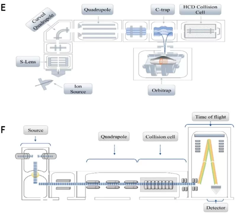
Figure 1. Orbitrap and Q-TOF configurations for high-resolution accurate-mass analysis. The Orbitrap panel illustrates ion transfer through the quadrupole and C-trap to Orbitrap mass analysis, whereas the Q-TOF panel illustrates quadrupole-based ion handling followed by time-of-flight mass analysis. Cropped from Figure 2 (panels E and F) in Guo et al. (2023), Frontiers in Chemistry, licensed under the Creative Commons Attribution 4.0 International License (CC BY 4.0). The image was cropped to retain panels E and F; no other content changes were made.
3. How Accurate Mass Narrows Metabolite Candidates
In metabolite annotation, one of the main practical benefits of accurate mass is reducing the number of plausible molecular formulas associated with a measured ion. The extent of that reduction depends on correct ion assignment, the mass-tolerance window, elemental constraints, isotope information, and the chemical context.
3.1 Mass-Error Windows for Candidate Filtering
At low resolution, many elemental compositions can collapse into the same nominal mass. HRMS allows candidates to be filtered within a narrow mass-error window around the measured value. A candidate outside that window can be excluded, whereas candidates within it remain possible. This simple step can substantially improve the selectivity of a metabolomics database search.
The window should not be chosen mechanically. A defensible tolerance reflects the observed calibration and quality-control performance of the data set, along with m/z range, ion intensity, acquisition mode, and instrument behavior. The number of surviving candidates also depends on which elements and adducts are allowed. Even extremely high mass accuracy does not guarantee a unique molecular formula or structure, especially as molecular mass and elemental complexity increase (Kind & Fiehn, 2006).
3.2 Molecular Formula Assignment with Isotope Patterns
Accurate mass becomes more informative when it is combined with chemical constraints. Candidate formula generation can incorporate allowed element ranges, isotope peak spacing and relative abundance, valence and double-bond-equivalent constraints, plausible H/C and O/C relationships, and, where appropriate, nitrogen-rule considerations. Ion mode and adduct assignment are equally important because a formula should explain the measured ion rather than an assumed neutral molecule.
Modern computational tools combine these constraints with isotope patterns and, when available, fragmentation data to rank molecular formulas. SIRIUS, for example, integrates isotope and tandem-MS information to support molecular formula and structure inference (Dührkop et al., 2019). When accurate mass is evaluated together with isotope patterns and chemical constraints, formula assignment generally produces a ranked set of plausible compositions rather than a single definitive formula.
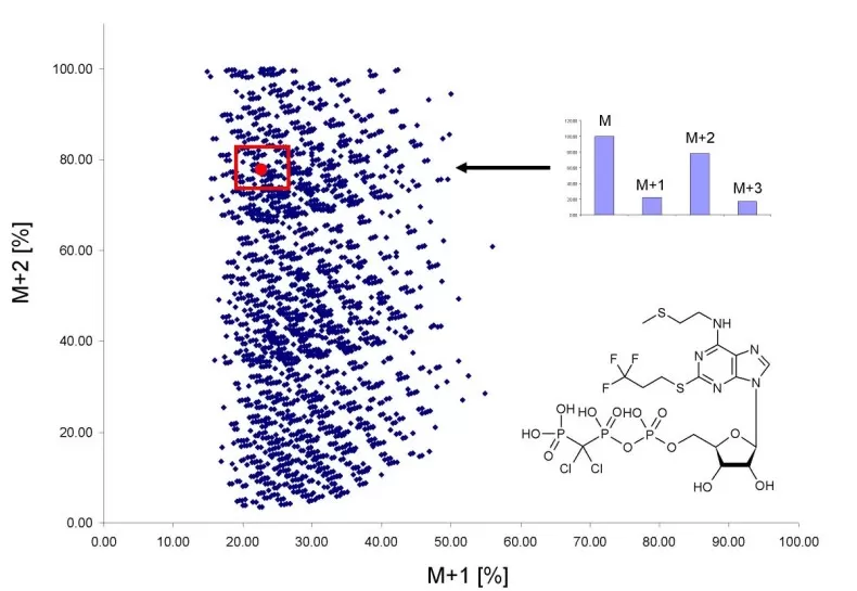
Figure 2. Accurate mass and isotope-pattern constraints reduce molecular-formula ambiguity. The plot shows the relative M+1 and M+2 isotope abundances of elemental compositions compatible with the measured mass of Cangrelor at 1 ppm mass accuracy. The red box marks formulas consistent with the ±5% isotope-ratio criterion used in this example, illustrating how isotope-abundance information can substantially reduce the number of mass-compatible formulas. Reproduced from Figure 7 in Kind and Fiehn (2007), BMC Bioinformatics, licensed under the Creative Commons Attribution 2.0 License (CC BY 2.0); no content changes were made.
3.3 Isobars vs. Isomers in HRMS
The distinction between isobars and structural isomers is critical when interpreting HRMS data. Isobaric or near-isobaric compounds can have very similar nominal masses while differing slightly in exact mass because their elemental compositions differ. If their m/z separation is sufficiently large relative to the peak width at that m/z, HRMS can resolve the signals as distinct peaks, while accurate mass can eliminate incompatible molecular formulas.
True structural isomers are different. Compounds with the same molecular formula have the same exact mass, so accurate mass alone cannot distinguish them. Consider a positive-mode feature near m/z 181.0707 assigned as [M+H]+. A neutral monoisotopic mass near 180.0634 is compatible with the formula C6H12O6 under appropriate constraints, but glucose, fructose, and galactose all share that formula. Their distinction requires orthogonal information such as chromatographic separation, diagnostic MS/MS, retention-time comparison, ion mobility/collision cross section, derivatization, or authentic standards. Stereoisomers can be even more demanding.
Accurate mass can therefore narrow many near-isobaric candidates, whereas structural isomer assignments require orthogonal evidence.
4. Accurate-Mass Database and Spectral Searching
Database searching becomes more informative once the ion form and candidate formula have been constrained. Structure databases, pathway resources, and spectral libraries serve different purposes, and no single resource is sufficient for every annotation task.
4.1 Structure, Pathway, and Context Databases
HMDB is particularly useful when human biological context matters because it links chemical information with human metabolite, biospecimen, pathway, and spectral information (Wishart et al., 2022). KEGG COMPOUND connects chemical entities with biochemical reactions and pathway maps, making it useful for evaluating whether a candidate is compatible with known metabolic context (Kanehisa et al., 2023).
Broader resources such as PubChem and ChEBI expand structural candidates and standardize identifiers, while LIPID MAPS is useful for lipid-specific nomenclature and structures. Biological plausibility can prioritize candidates, but it should not override contradictory analytical evidence.
4.2 MS/MS Spectral Libraries
Accurate precursor mass generates candidates, while MS/MS libraries add structural evidence. MassBank provides searchable reference spectra for small molecules (Horai et al., 2010); MoNA aggregates records from multiple sources; GNPS combines community spectral libraries with library search and molecular networking (Wang et al., 2016); and METLIN provides metabolite and tandem-MS resources.
Collision energy, instrument type, precursor isolation, ion mode, adduct, and library provenance can alter fragmentation. Experimental and predicted spectra should be distinguished, and a high match score is not equivalent to confirmation with a matched authentic standard.
Table 2. Databases and Spectral Resources for HRMS Metabolite Annotation
| Resource(s) | Resource type | Primary use | Evidence contribution | Main caution |
|---|---|---|---|---|
| HMDB | Human metabolite and biological-context database | Prioritize candidates in human samples | Formula, structures, names, biological context, selected spectra | Human-centered; absence does not prove absence from a sample |
| KEGG COMPOUND | Biochemical compound and pathway resource | Connect candidates with reactions and pathways | Formula, structure, reaction/pathway context | Pathway membership does not confirm identity |
| PubChem / ChEBI | Broad chemical structure database / chemical ontology | Expand structures, synonyms, identifiers, and classes | Formula, structure, identifiers, ontology | Broad candidate space; not spectral confirmation |
| LIPID MAPS | Lipid-specialized resource | Generate lipid-class and lipid-structure candidates | Formula, exact mass, lipid nomenclature and structures | Annotation level must match the evidence available |
| MassBank / MoNA | MS/MS spectral libraries and aggregators | Match experimental spectra to references | Reference fragmentation patterns and metadata | Conditions and metadata can be heterogeneous |
| GNPS | Community mass-spectrometry platform | Library search, dereplication, molecular networking | MS/MS similarity and community-curated resources | Coverage is incomplete and provenance matters |
| METLIN | Metabolite and MS/MS resource | Accurate-mass and tandem-MS candidate search | Chemical records and spectral information | Access, evidence type, and experimental conditions should be checked |
4.3 From Molecular Formula to Structural Candidates
A reproducible search should record ion/adduct assignment, neutral monoisotopic mass or formula, mass tolerance, database or spectral resource, version or access date when relevant, and sample-specific filters. Synonyms and duplicate identifiers should be normalized. Species, tissue, exposure, pathway, and compound class can then prioritize candidates, but several same-formula structures may remain valid. The first database hit is therefore a candidate, not a confirmed identity.
5. Evidence Beyond Accurate Mass for Metabolite Identification
Accurate precursor mass primarily constrains elemental composition and candidate space. MS/MS fragments and neutral losses can add evidence for substructures, functional groups, and compound classes, while chromatographic retention, ion mobility-derived collision cross section (CCS), and authentic standards can strengthen discrimination among isomeric candidates. Computational tools can also use fragmentation patterns to rank structures, but their output remains dependent on the spectra, candidate database, and model assumptions (Dührkop et al., 2019; Alseekh et al., 2021).
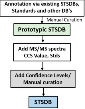
Figure 3. Orthogonal evidence for higher-confidence metabolite annotation. This cropped sample type–specific database (STSDB) workflow shows MS/MS spectra, collision cross section (CCS) values, and authentic-standard data being added before confidence-level curation. Cropped from Figure 3 in Reisdorph et al. (2019), Metabolites, licensed under CC BY 4.0; no other content changes were made.
For transparent reporting, important features should retain the observed m/z, ion/adduct assignment, mass error, candidate formula, database or spectral resource, MS/MS evidence, and any orthogonal confirmation. Reporting these evidence layers together makes the basis for each annotation explicit and helps distinguish tentative annotations from stronger identifications.
Related reading: Metabolite Identification in LC-MS Metabolomics: Identification Principles and Confidence Levels
6. Common HRMS Annotation Pitfalls
Several recurring mistakes can make high-resolution data appear more conclusive than they are:
- Terminology and ion assignment: Do not use accurate mass and exact mass interchangeably, and do not treat observed m/z as the neutral molecular mass without considering charge and adduct form.
- Unjustified search settings: Mass tolerance, elemental ranges, and allowed adducts should be supported by calibration, QC performance, sample chemistry, and acquisition conditions.
- Overinterpreting candidate rankings: A top database result or spectral score is a prioritized hypothesis unless additional evidence supports the structure.
- Confusing isobars with isomers: HRMS can resolve many near-isobaric signals but cannot distinguish true structural or stereochemical isomers by accurate mass alone.
- Incomplete evidence reporting: Database version, search parameters, experimental versus predicted spectra, retention information, CCS, and standard comparisons should be reported when they influence the claim.
Reliable annotation depends on a transparent, mutually consistent combination of mass accuracy, spectral evidence, and orthogonal measurements.
HRMS Metabolite Identification FAQs
Is accurate mass the same as exact mass?
No. Accurate mass is an experimentally measured ion mass, whereas exact mass is a calculated value for a specified isotopic composition. The distinction is important when comparing measured m/z values with theoretical formulas (Murray et al., 2013).
Can Orbitrap or Q-TOF distinguish metabolite isomers?
Not by accurate mass alone. Orbitrap and Q-TOF can separate many near-isobaric ions that overlap at lower resolution, but true isomers with the same molecular formula share the same exact mass. LC separation, diagnostic MS/MS, CCS, derivatization, or authentic standards may therefore be required.
Which database or spectral resource should be used for accurate-mass searching?
No single resource is optimal for every sample. HMDB is useful for human biological context, KEGG for pathway-linked candidates, PubChem and ChEBI for broad structural space, LIPID MAPS for lipids, and MassBank, MoNA, GNPS, or METLIN for tandem-MS comparison. Multiple resources are often complementary rather than interchangeable.
High-Resolution LC-MS Metabolomics at MetwareBio
A high-resolution metabolomics workflow spans acquisition, quality control, feature processing, annotation, and biological interpretation. MetwareBio supports untargeted metabolomics using high-resolution LC-MS/MS, with downstream feature processing, database-assisted annotation, statistical analysis, and pathway interpretation. Selected features can be followed by additional MS/MS evaluation or targeted verification when higher identification confidence is required.
Planning an untargeted metabolomics study or evaluating the identity of important metabolic features? Contact MetwareBio to discuss a high-resolution LC-MS/MS and annotation strategy matched to the sample type, biological question, and required identification confidence.
Contact UsRead More: Mass Spectrometry and Metabolomics Annotation
Explore these related articles to deepen your understanding of mass spectrometry platforms, metabolomics workflows, and data processing strategies that complement high-resolution accurate-mass analysis.
Compare the leading mass spectrometry platforms side by side, including Orbitrap, Q-TOF, and triple quadrupole systems. This guide helps you understand how resolving power, mass accuracy, scan speed, and sensitivity differ across instruments for metabolomics and proteomics applications.
Understand the fundamental differences between LC-MS and GC-MS platforms, including ionization methods, analyte coverage, and sample requirements. This comparison is essential for selecting the right mass spectrometry approach for your metabolomics study.
Learn how targeted, untargeted, and widely-targeted metabolomics strategies differ in scope, sensitivity, and annotation depth. This article provides practical guidance for choosing the approach that best matches your research question and identification confidence requirements.
Walk through the complete untargeted metabolomics workflow from sample preparation through feature detection, alignment, annotation, and biological interpretation. This guide complements high-resolution mass spectrometry by contextualizing accurate-mass data within the full analytical pipeline.
Explore the principles of electrospray ionization, the most widely used ionization technique in LC-MS metabolomics. Understanding ESI helps interpret adduct formation, charge states, and ion suppression effects that directly affect accurate-mass measurement and metabolite annotation.
Discover advanced data processing methods for metabolomics, including feature alignment, gap filling, batch correction, and statistical filtering. These techniques are essential for converting high-resolution raw data into reliable feature tables for accurate-mass database searching and annotation.
References
- Alseekh, S., Aharoni, A., Brotman, Y., et al. (2021). Mass spectrometry-based metabolomics: A guide for annotation, quantification and best reporting practices. Nature Methods, 18, 747-756. https://doi.org/10.1038/s41592-021-01197-1
- Chernushevich, I. V., Loboda, A. V., & Thomson, B. A. (2001). An introduction to quadrupole-time-of-flight mass spectrometry. Journal of Mass Spectrometry, 36(8), 849-865. https://doi.org/10.1002/jms.207
- Dührkop, K., Fleischauer, M., Ludwig, M., et al. (2019). SIRIUS 4: A rapid tool for turning tandem mass spectra into metabolite structure information. Nature Methods, 16, 299-302. https://doi.org/10.1038/s41592-019-0344-8
- Guo, S., Qiu, S., Cai, Y., Wang, Z., Yang, Q., Tang, S., Xie, Y., & Zhang, A. (2023). Mass spectrometry-based metabolomics for discovering active ingredients and exploring action mechanism of herbal medicine. Frontiers in Chemistry, 11, 1142287. https://doi.org/10.3389/fchem.2023.1142287
- Horai, H., Arita, M., Kanaya, S., et al. (2010). MassBank: A public repository for sharing mass spectral data for life sciences. Journal of Mass Spectrometry, 45(7), 703-714. https://doi.org/10.1002/jms.1777
- Kanehisa, M., Furumichi, M., Sato, Y., Kawashima, M., & Ishiguro-Watanabe, M. (2023). KEGG for taxonomy-based analysis of pathways and genomes. Nucleic Acids Research, 51(D1), D587-D592. https://doi.org/10.1093/nar/gkac963
- Kind, T., & Fiehn, O. (2006). Metabolomic database annotations via query of elemental compositions: Mass accuracy is insufficient even at less than 1 ppm. BMC Bioinformatics, 7, 234. https://doi.org/10.1186/1471-2105-7-234
- Kind, T., & Fiehn, O. (2007). Seven Golden Rules for heuristic filtering of molecular formulas obtained by accurate mass spectrometry. BMC Bioinformatics, 8, 105. https://doi.org/10.1186/1471-2105-8-105
- Makarov, A. (2000). Electrostatic axially harmonic orbital trapping: A high-performance technique of mass analysis. Analytical Chemistry, 72(6), 1156-1162. https://doi.org/10.1021/ac991131p
- Murray, K. K., Boyd, R. K., Eberlin, M. N., Langley, G. J., Li, L., & Naito, Y. (2013). Definitions of terms relating to mass spectrometry (IUPAC Recommendations 2013). Pure and Applied Chemistry, 85(7), 1515-1609. https://doi.org/10.1351/PAC-REC-06-04-06
- Reisdorph, N. A., Walmsley, S., & Reisdorph, R. (2019). A perspective and framework for developing sample type specific databases for LC/MS-based clinical metabolomics. Metabolites, 10(1), 8. https://doi.org/10.3390/metabo10010008
- Wang, M., Carver, J. J., Phelan, V. V., et al. (2016). Sharing and community curation of mass spectrometry data with Global Natural Products Social Molecular Networking. Nature Biotechnology, 34, 828-837. https://doi.org/10.1038/nbt.3597
- Wishart, D. S., Guo, A., Oler, E., et al. (2022). HMDB 5.0: The Human Metabolome Database for 2022. Nucleic Acids Research, 50(D1), D622-D631. https://doi.org/10.1093/nar/gkab1062